. 2007 May 22;104(21):9035-40.
doi: 10.1073/pnas.0703285104. Epub 2007 May 15.

Double-stranded DNA and double-stranded RNA induce a common antiviral signaling pathway in human cells

Affiliations

Double-stranded DNA and double-stranded RNA induce a common antiviral signaling pathway in human cells

Guofeng Cheng et al. Proc Natl Acad Sci U S A. .

Abstract

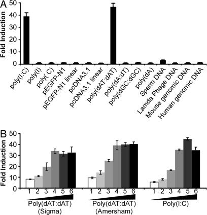
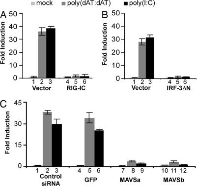
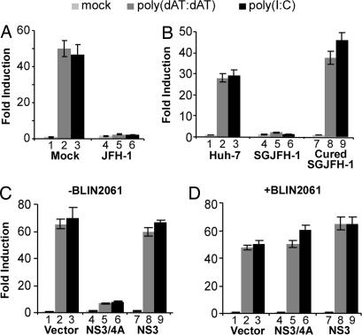
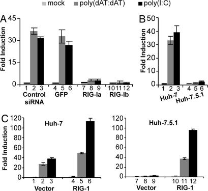
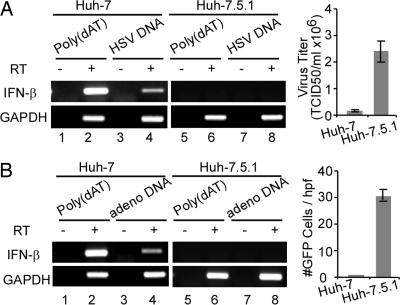
Virus infection triggers IFN immune defenses in infected cells in part through viral nucleic acid interactions, but the pathways by which dsDNA and DNA viruses trigger innate defenses are only partially understood. Here we present evidence that both retinoic acid-induced gene I (RIG-I) and mitochondrial antiviral signaling protein (MAVS) are required for dsDNA-induced IFN-beta promoter activation in a human hepatoma cell line (Huh-7), and that activation is efficiently blocked by the hepatitis C virus NS3/4A protease, which is known to block dsRNA signaling by cleaving MAVS. These findings suggest that dsDNA and dsRNA share a common pathway to trigger the innate antiviral defense response in human cells, although dsDNA appears to trigger that pathway upstream of the dsRNA-interacting protein RIG-I.


Conflict of interest statement

The authors declare no conflict of interest.

Figures

Fig. 1.
Fig. 1.
Cytosolic dsDNA activates the IFN-β promoter in Huh-7 cells. (A) Huh-7 cells were first cotransfected with the IFN-β promoter luciferase reporter plasmid, an internal control plasmid pRL-TK, and a carrier plasmid pcDNA3.1. At 36 h after transfection, cells were either mock-transfected or transfected with various DNA or RNA stimuli (1.0 μg/ml) using Lipofectamine 2000. Cells were subjected to dual luciferase assay 16 h after transfection. The results are expressed as fold induction of IFN-β promoter activity relative to the basal level. (B) Dose titration of dsDNA and dsRNA to induce IFN-β promoter activation. The experimental conditions were the same as in A except that the indicated amount of dsDNA or dsRNA was transfected. (1-6: 0.125, 0.25, 0.5, 1.0, 2.0, and 4.0 μg/ml).
Fig. 2.
Fig. 2.
MAVS is essential for dsDNA-induced IFN-β promoter activation. (A and B) RIG-I and IRF-3 dominant-negative mutants block dsDNA-induced IFN-β promoter activation. Huh-7 cells were cotransfected with the IFN-β promoter luciferase reporter and plasmid pRL-TK in the presence of an empty vector or a plasmid expressing RIG-IC or IRF-3 ΔN. Thirty-six hours later, cells were mock-transfected or transfected with poly(dAT:dAT) or poly(I:C). Luciferase activities were measured 16 h after transfection. (C) Knockdown of MAVS blocks dsDNA-induced IFN-β promoter activation. siRNAs were transfected into Huh-7 cells as described in Material and Methods. At 24 h after transfection, cells were transfected with the IFN-β promoter luciferase reporter and subjected to the luciferase reporter assay.
Fig. 3.
Fig. 3.
HCV NS3/4A blocks dsDNA-induced IFN-β promoter activation. (A) HCV infection blocks dsDNA-induced IFN-β promoter activation. Huh-7 cells were either mock-infected or infected with JFH-1 at a multiplicity of infection of 1. On day 4 after infection, cells were transfected with the IFN-β promoter luciferase reporter and subjected to the luciferase reporter assay. (B) dsDNA-induced IFN-β promoter activation is blocked in the HCV replicon cells. Parental Huh-7 cells, JFH-1 subgenomic replicon cells (SGJFH-1) and cured replicon cells were transfected with the IFN-β promoter luciferase reporter and subjected to the luciferase reporter assay. (C) HCV NS3/4A protease blocks dsDNA-induced IFN-β promoter activation. Huh-7 cells were cotransfected with the IFN-β promoter luciferase reporter and plasmid pRL-TK in the presence of an empty vector or a plasmid expressing either NS3 or NS3/4A. 36 h later, the IFN-β luciferase activity induced by poly(dAT:dAT) or poly(I:C) was assayed as described in Materials and Methods. (D) BILN2061 prevents the blocking activity of NS3/4A. The experimental conditions were the same as in C, except that the protease inhibitor BILN2061 was added to the cells 1 h before plasmid transfection at a final concentration of 5 μM.
Fig. 4.
Fig. 4.
RIG-I is essential for the dsDNA-induced IFN-β promoter activation. (A) Knockdown of RIG-I blocks dsDNA-induced IFN-β promoter activation. siRNAs were transfected into Huh-7 cells as described in Materials and Methods. At 24 h after transfection, cells were transfected with the IFN-β promoter luciferase reporter and subjected to the luciferase reporter assay. (B) dsDNA-induced IFN-β promoter activation is absent in Huh-7.5.1 cells. Huh-7 and Huh-7.5.1 cells were transfected with the IFN-β promoter luciferase reporter and the luciferase activity induced by poly(dAT:dAT) or poly(I:C) was assayed as described in Materials and Methods. (C) Wild-type RIG-I restores dsDNA-induced IFN-β promoter activation in Huh-7.5.1 cells. Huh-7 (Left) or Huh-7.5.1 (Right) cells were cotransfected with the IFN-β promoter luciferase reporter and plasmid pRL-TK in the presence of an empty vector or a plasmid expressing wild-type RIG-I. The IFN-β luciferase activity induced by poly(dAT:dAT) or poly(I:C) was assayed as in B.
Fig. 5.
Fig. 5.
Huh-7.5.1 cells are more permissive to DNA virus infections. (A) HSV-1 infection is more productive in Huh-7.5.1 cells than in Huh-7 cells. (Left) Huh-7 and Huh-7.5.1 cells were transfected with purified HSV-1 DNA at a concentration of 10 μg/ml by using lipofectamine 2000. At 18 h after transfection, cells were harvested and the transcriptional levels of IFN-β and GAPDH were analyzed by using RT-PCR described in Material and Methods. RT reactions without reverse transcriptase were used for DNA contamination control. (Right) Huh-7 and Huh-7.5.1 cells were infected with HSV-1 at multiplicity of infection of 0.05. At 24 h after infection, HSV-1-infected cells were harvested and virus titers were determined in Vero cells. (B) Adenovirus replicates more efficiently in Huh-7.5.1 cells. The DNA transfection and adenovirus infection were done similarly as described in A, except that adenovirus infection was measured by the number of GFP-expressing cells.

References

    1. Akira S, Uematsu S, Takeuchi O. Cell. 2006;124:783–801. - PubMed
    1. Kawai T, Akira S. Curr Opin Immunol. 2005;17:338–344. - PubMed
    1. Yoneyama M, Kikuchi M, Natsukawa T, Shinobu N, Imaizumi T, Miyagishi M, Taira K, Akira S, Fujita T. Nat Immunol. 2004;5:730–737. - PubMed
    1. Yoneyama M, Kikuchi M, Matsumoto K, Imaizumi T, Miyagishi M, Taira K, Foy E, Loo YM, Gale M, Jr, Akira S, et al. J Immunol. 2005;175:2851–2858. - PubMed
    1. Hiscott J, Lin R, Nakhaei P, Paz S. Trends Mol Med. 2006;12:53–56. - PubMed

Publication types

MeSH terms