. 2007 May 29;104(22):9209-12.
doi: 10.1073/pnas.0703337104. Epub 2007 May 21.

High-resolution scanning tunneling microscopy imaging of mesoscopic graphene sheets on an insulating surface

Affiliations

High-resolution scanning tunneling microscopy imaging of mesoscopic graphene sheets on an insulating surface

Elena Stolyarova et al. Proc Natl Acad Sci U S A. .

Abstract

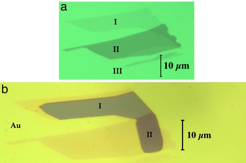
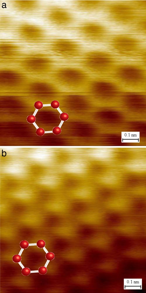
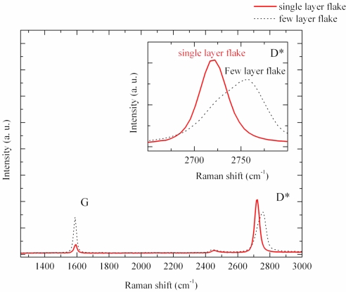
We present scanning tunneling microscopy (STM) images of single-layer graphene crystals examined under ultrahigh vacuum conditions. The samples, with lateral dimensions on the micrometer scale, were prepared on a silicon dioxide surface by direct exfoliation of crystalline graphite. The single-layer films were identified by using Raman spectroscopy. Topographic images of single-layer samples display the honeycomb structure expected for the full hexagonal symmetry of an isolated graphene monolayer. The absence of observable defects in the STM images is indicative of the high quality of these films. Crystals composed of a few layers of graphene also were examined. They exhibited dramatically different STM topography, displaying the reduced threefold symmetry characteristic of the surface of bulk graphite.


Conflict of interest statement

The authors declare no conflict of interest.

Figures

Fig. 1.
Fig. 1.
Optical microscopy images of the graphene flake examined in this study. (a) An image of the sample before deposition of the electrode. Three regions with different optical densities can be identified: I, single-layer graphene; II, multilayer graphene; and III, the silicon-dioxide-coated substrate. (b) An image of the same flake after the deposition of an 18-nm layer of gold. The gold electrode completely covers the substrate and partially covers the graphitic flake. The darker region is the uncovered part.
Fig. 2.
Fig. 2.
STM topographic images of different regions of the graphene flake of Fig. 1. The images were obtained with Vbias = +1 V (sample potential), I = 1 nA, and a scan area of 1 nm2. A model of the underlying atomic structure is shown as a guide to the eye. (a) Image from a single layer of graphene (region I of Fig. 1). A honeycomb structure is observed. (b) Image of the multilayer portion of the sample (region II of Fig. 1). The characteristic three-for-six STM image of the surface of bulk graphite is observed.
Fig. 3.
Fig. 3.
Stereographic plot of a large-scale (100 × 62 nm) STM image of a single-layer graphene film on the silicon dioxide surface. The STM scanning conditions were Vbias = 1 V (sample potential) and I = 0.6 nA. The 0.8-nm scale of the vertical (Z) coordinate is greatly enlarged to accentuate the surface features.
Fig. 4.
Fig. 4.
Comparison of Raman spectra at 457.5 nm for single-layer (solid line) and multilayer (dashed line) regions of the graphitic flake described in Fig. 1. The two intense features are the G peak at a Raman shift of ≈1,580 cm−1 and the D* band at ≈2,710 cm−1. The D* band (enlarged in Inset) of a few-layer flake is blue-shifted and broadened with respect to that of the single-layer graphene sample. Moreover, the D* peak of single-layer graphene is symmetric, whereas the D* band corresponding to the multilayer sample has a complex asymmetric shape.

References

    1. Novoselov KS, Geim AK, Morozov SV, Jiang D, Zhang Y, Dubonos SV, Grigorieva IV, Firsov AA. Science. 2004;306:666–669. - PubMed
    1. Heersche HB, Jarillo-Herrero P, Oostinga JB, Vandersypen LMK, Morpurgo AF. Nature. 2007;446:56–59. - PubMed
    1. Zhang Y, Jiang Z, Small JP, Purewal MS, Tan YW, Fazlollahi M, Chudow JD, Jaszczak JA, Stormer HL, Kim P. Phys Rev Lett. 2006;96:136806. - PubMed
    1. Zhang YB, Tan YW, Stormer HL, Kim P. Nature. 2005;438:201–204. - PubMed
    1. Novoselov KS, Geim AK, Morozov SV, Jiang D, Katsnelson MI, Grigorieva IV, Dubonos SV, Firsov AA. Nature. 2005;438:197–200. - PubMed

LinkOut - more resources