. 2007 May 22;104(21):8773-8.
doi: 10.1073/pnas.0701124104. Epub 2007 May 16.

Structure of the Munc18c/Syntaxin4 N-peptide complex defines universal features of the N-peptide binding mode of Sec1/Munc18 proteins

Affiliations

Structure of the Munc18c/Syntaxin4 N-peptide complex defines universal features of the N-peptide binding mode of Sec1/Munc18 proteins

Shu-Hong Hu et al. Proc Natl Acad Sci U S A. .

Abstract

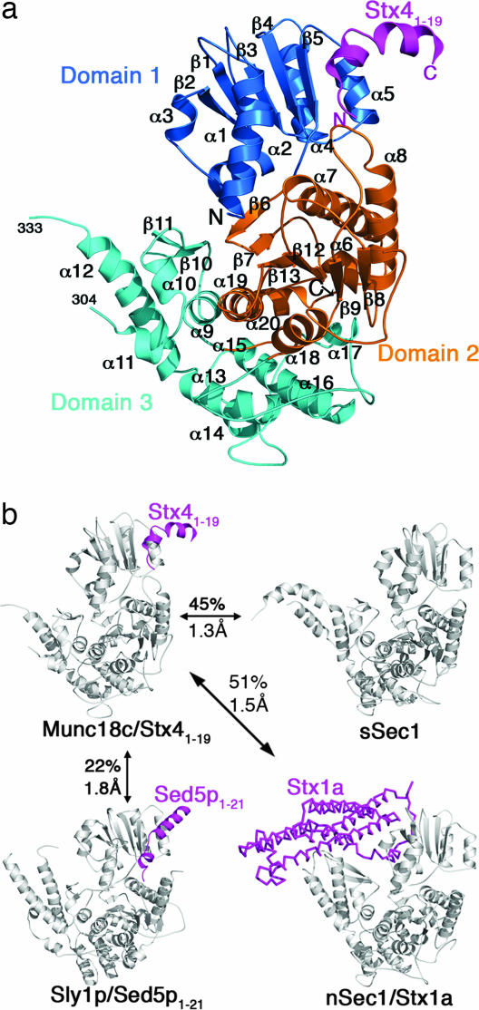
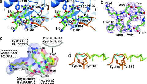
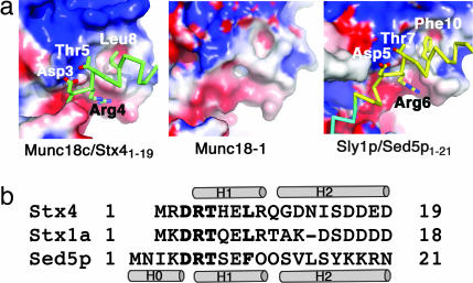
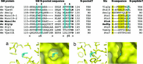
Sec1/Munc18 proteins (SM proteins) bind to soluble NSF attachment protein receptors (SNAREs) and play an essential role in membrane fusion. Divergent modes of regulation have been proposed for different SM proteins indicating that they can either promote or inhibit SNARE assembly. This is in part because of discrete modes of binding that have been described for various SM/SNARE complexes. One mode suggests that SM proteins bind only to Syntaxins (Stx) preventing SNARE assembly, whereas in another they facilitate SNARE assembly and bind to SNARE complexes. The mammalian cell surface SM protein Munc18c binds to an N-peptide in Stx4, and this is compatible with its interaction with SNARE complexes. Here we describe the crystal structure of Munc18c in complex with the Stx4 N-peptide. This structure shows remarkable similarity with a yeast complex indicating that the mode of binding, which can accommodate SNARE complexes, is highly conserved throughout evolution. Modeling reveals the presence of the N-peptide binding mode in most but not all yeast and mammalian SM/Stx pairs, suggesting that it has coevolved to fulfill a specific regulatory function. It is unlikely that the N-peptide interaction alone accounts for the specificity in SM/SNARE binding, implicating other contact surfaces in this function. Together with other data, our results support a sequential two-state model for SM/SNARE binding involving an initial interaction via the Stx N-peptide, which somehow facilitates a second, more comprehensive interaction comprising other contact surfaces in both proteins.


Conflict of interest statement

The authors declare no conflict of interest.

Figures

Fig. 1.
Fig. 1.
Structure of Munc18c/Stx41–19 and comparison with other SM proteins. (a) The Munc18c/Stx41–19 complex showing domain 1 (residues 1–138) in dark blue, domain 2 (residues 139–250 and 476–584) in orange, and domain 3 (residues 251–475) in cyan. Stx4 residues 1–19 are shown in magenta. Helices are shown as coils, and strands are shown as arrows. N and C termini and secondary structure elements are labeled. (b) Comparison of mouse Munc18c/Stx41–19 with squid neuronal Munc18-1 [sSec1, PDB ID code 1EPU (23)], yeast Sly1p/Sed5p1–21 [PDB ID code 1MQS (20)], and rat neuronal Munc18-1/Stx1a (nSec1/Stx1a) [PDB ID code 1DN1 (10)] showing rmsd of the structures and the pairwise sequence identity.
Fig. 2.
Fig. 2.
Interaction between Stx4 N-peptide and Munc18c. (a) Stereo diagram showing the bound N-peptide (green) and residues of Munc18c (blue, domain 1; orange, domain 2). Hydrogen bond interactions are shown as dotted lines. (b) Electron density of the Stx4 N-peptide (2FoFc contoured at 1σ). (c) Superimposition of SM-bound conformations of Stx4 (green) and Sed5p (yellow) N-peptides. Intramolecular hydrogen bonds (blue dotted lines) and Munc18c-interacting residues are indicated (hydrogen bond partners for Arg-2, Asp-3, Arg-4, and Thr-5 and hydrophobic interacting partners for Leu-8). The electrostatic surface for the Stx4 peptide is also shown, revealing a basic face (Arg-2 and Arg-4) and an acidic face (Asp-3 and Glu-7) that are complementary to the Munc18c binding surface (Fig. 3). (d) Stereo diagram of the α8 loop region of Munc18c (orange) and the Stx4 (green) and Sed5p (yellow and blue) N-peptides, showing the overlap between the loop and the nonnative residues of Sed5p (blue).
Fig. 3.
Fig. 3.
A structurally conserved N-peptide binding site in Munc18c, Sly1p, and Munc18-1. (a) Electrostatic surfaces are shown for the structures of Munc18c, Munc18-1, and Sly1p in the N-peptide binding region (colored blue to red from +5 to −5 KbT/e). An acidic groove (red), basic patch (blue), and hydrophobic pocket (white) characterize the N-peptide binding sites of Munc18c and Sly1p, and these features are also present in Munc18-1. The main chain of the N-peptides of Stx4 (green) and Sed5p (yellow and blue) and the side chains of the DRT motif and hydrophobic residues are shown. (b) Sequence alignment of the N-peptides of mouse Stx4, rat Stx1a, and yeast Sed5p is shown with the DRT motif and hydrophobic residue indicated in bold. Helices are shown as cylinders for Stx4 and Sed5p.
Fig. 4.
Fig. 4.
SM proteins predicted to have a hydrophobic N-pocket have cognate Stxs with an N-peptide sequence motif. A sequence alignment of the N-pocket of SM proteins from yeast (S. cerevisiae, Sc) and mammals (M. musculus, Mm) with the proteins involved in the two N-peptide complexes that have been structurally characterized shown in bold. Hydrophobic residues that align with sites 1, 2, 3, and 4 of the N-pockets of Munc18c and Sly1p are highlighted in cyan. The SM protein acidic residue that interacts with the DRT motif of the N-peptide is shown in bold. All SM proteins except Sec1p, Vps33p, Vps33A, and Vps33B (data not shown) are predicted to have hydrophobic N-pockets that could interact with the N-peptide hydrophobic residue of cognate Stxs. All Stxs except the cognate Stxs of Sec1p, Vps33p, and Vps33A/B have an N-peptide motif (shown in yellow with the hydrophobic residue in cyan). For the Stx N-peptides, alignment of the N-terminal sequences of Sso1p and Vam3p is based on the sequence alignment in ref. . (a) Munc18c residues from the N-pocket (yellow) that interact with the hydrophobic residue of the Stx4 N-peptide Leu-8 (cyan). The surface of the hydrophobic N-pocket is also shown. (b) The same region is shown for yeast Sec1p, predicted from the structure of Munc18c and the sequence alignment above, showing that the hydrophobic residues at sites 1–4 of Sec1p fill the pocket.

References

    1. Bennett MK, Garcia-Arraras JE, Elferink LA, Peterson K, Fleming AM, Hazuka CD, Scheller RH. Cell. 1993;74:863–873. - PubMed
    1. Brunger AT. Q Rev Biophys. 2005;38:1–47. - PubMed
    1. Sutton RB, Fasshauer D, Jahn R, Brunger AT. Nature. 1998;395:347–353. - PubMed
    1. Rizo J, Sudhof TC. Nat Rev Neurosci. 2002;3:641–653. - PubMed
    1. Jahn R, Scheller RH. Nat Rev Mol Cell Biol. 2006;7:631–643. - PubMed

Publication types

MeSH terms

Associated data