

fMRI investigation of working memory for faces in autism: visual coding and underconnectivity with frontal areas
- PMID: 17517680
- PMCID: PMC4500154
- DOI: 10.1093/cercor/bhm054
fMRI investigation of working memory for faces in autism: visual coding and underconnectivity with frontal areas
Abstract
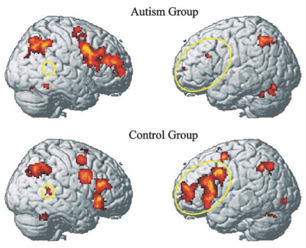
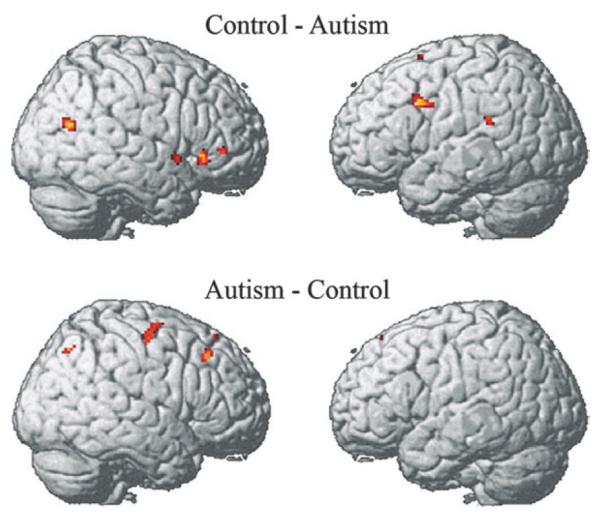
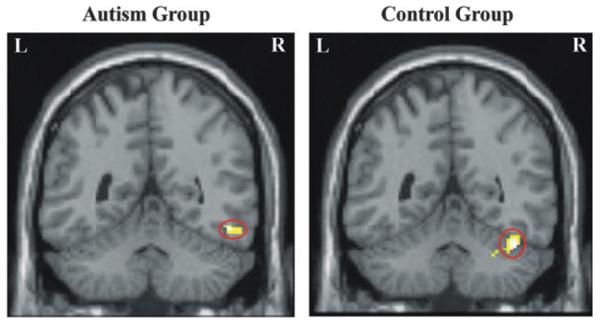
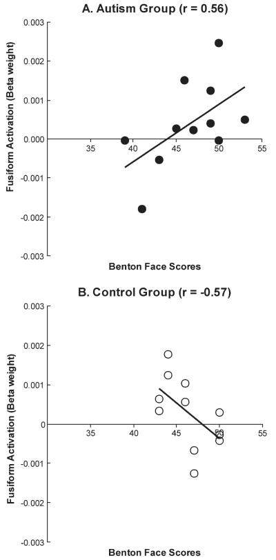
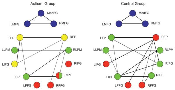
Brain activation and functional connectivity were investigated in high functioning autism using functional magnetic resonance imaging in an n-back working memory task involving photographic face stimuli. The autism group showed reliably lower activation compared with controls in the inferior left prefrontal area (involved in verbal processing and working memory maintenance) and the right posterior temporal area (associated with theory of mind processing). The participants with autism also showed activation in a somewhat different location in the fusiform area than the control participants. These results suggest that the neural circuitry of the brain for face processing in autism may be analyzing the features of the face more as objects and less in terms of their human significance. The functional connectivity results revealed that the abnormal fusiform activation was embedded in a larger context of smaller and less synchronized networks, particularly indicating lower functional connectivity with frontal areas. In contrast to the underconnectivity with frontal areas, the autism group showed no underconnectivity among posterior cortical regions. These results extend previous findings of abnormal face perception in autism by demonstrating that the abnormalities are embedded in an abnormal cortical network that manages to perform the working memory task proficiently, using a visually oriented, asocial processing style that minimizes reliance on prefrontal areas.
Figures
References
-
- Allison T, Puce A, McCarthy G. Social perception from visual cues: role of the STS region. Trends Cogn Sci. 2000;4:267–278. - PubMed
-
- Baddeley A. Working memory. Oxford University Press; New York: 1986.
-
- Barnea-Goraly N, Kwon H, Menon V, Eliez S, Lotspeich L, Reiss AL. White matter structure in autism: preliminary evidence from diffusion tensor imaging. Biol Psychiatry. 2004;55:323–326. - PubMed
-
- Baron-Cohen S, Ring HA, Wheelwright S, Bullmore ET, Brammer MJ, Simmons A, Williams SCR. Social intelligence in the normal and autistic brain: an fMRI study. Eur J Neurosci. 1999;11:1891–1898. - PubMed
-
- Basser PJ. Inferring microstructural features and the physiological state of tissues from diffusion-weighted images. NMR Biomed. 1995;8:333–344. - PubMed
