. 2007;35(11):3774-83.
doi: 10.1093/nar/gkm336. Epub 2007 May 21.

Three-dimensional model for the isolated recombinant influenza virus polymerase heterotrimer

Affiliations

Three-dimensional model for the isolated recombinant influenza virus polymerase heterotrimer

Eva Torreira et al. Nucleic Acids Res. 2007.

Abstract

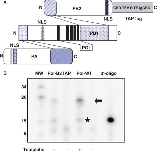
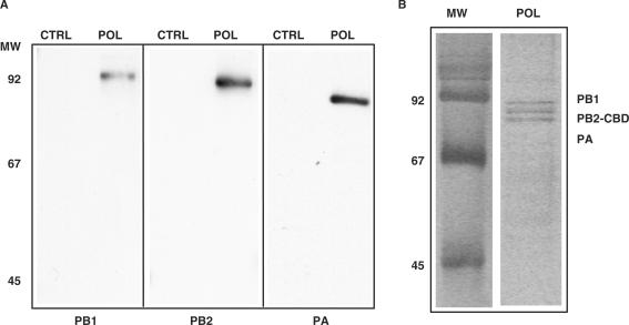
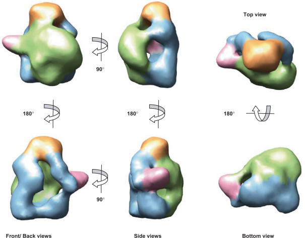
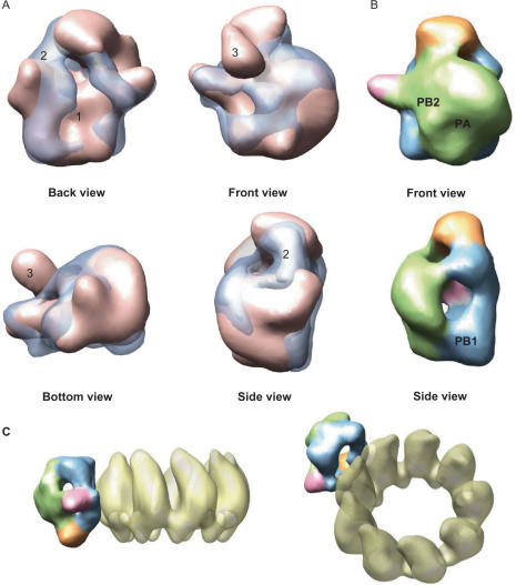
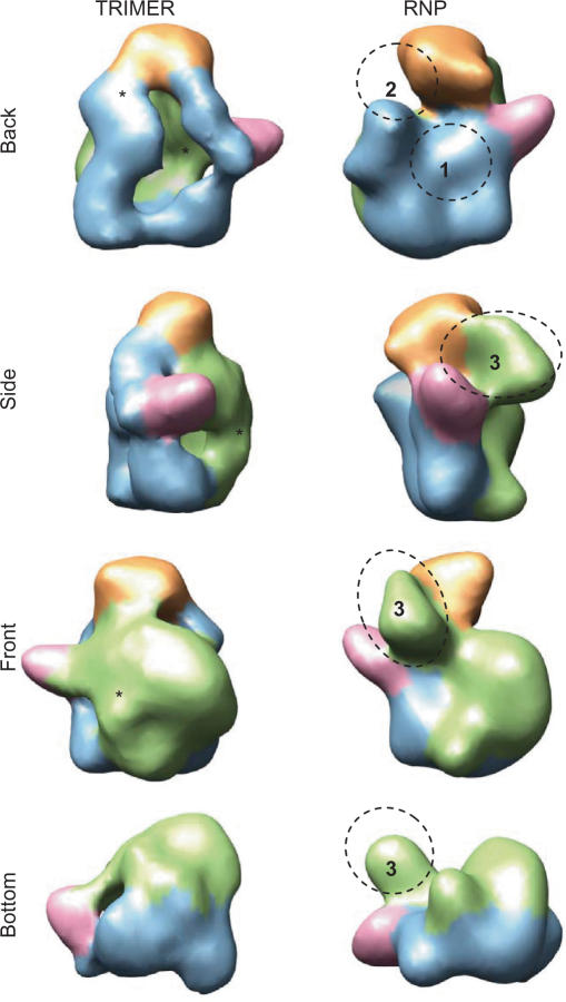
The genome of influenza A virus is organized into eight ribonucleoprotein complexes (RNPs), each containing one RNA polymerase complex. This RNA polymerase has also been found non-associated to RNPs and is possibly involved in distinct functions in the infection cycle. We have expressed the virus RNA polymerase complex by co-tranfection of the PB1, PB2 and PA genes in mammalian cells and the heterotrimer was purified by the TAP tag procedure. Its 3D structure was determined by electron microscopy and single-particle image processing. The model obtained resembles the structure previously reported for the polymerase complex associated to viral RNPs but appears to be in a more open conformation. Detailed model comparison indicated that specific areas of the complex show important conformational changes as compared to the structure for the RNP-associated polymerase, particularly in regions known to interact with the adjacent NP monomers in the RNP. Also, the PB2 subunit seems to undergo a substantial displacement as a result of the association of the polymerase to RNPs. The structural model presented suggests that a core conformation of the polymerase in solution exists but the interaction with other partners, such as proteins or RNA, will trigger distinct conformational changes to activate new functional properties.


Figures

Figure 1.
Figure 1.
In vitro activity of TAP-tagged polymerase. (A) Diagram showing the general organization of the polymerase complex including interaction domains (52–56), the position of the subunit NLSs (57–60) and the polymerase motifs within PB1 (61). Also shown is the structure of the TAP tag, fused to the C-terminus of PB2, with the calmodulin-binding domain (CBD), the TEV cleavage site and the IgG-binding domain (IgGBD). (B) The RNA synthesis activity of wild-type (Pol-WT) or TAP-tagged (Pol-B2TAP) polymerase-containing cell extracts was determined in vitro in the presence (+) or absence (−) of panhandle template, as indicated under the Materials and Methods section. The reaction products were analysed on a 18% polyacrylamide–urea gel along with molecular weight markers (MW), whose length in nt are indicated to the left. The 3′-terminal oligonucleotide present in the panhandle (3′-oligo) was terminally labelled with polynucleotide kinase and loaded in parallel as a size marker. The arrow indicates the position of the cap-snatched transcript and the star shows the position of the ApG-dependent transcript.
Figure 2.
Figure 2.
Purification of soluble polymerase complexes. (A)Western blot analyses of purified polymerase complex (POL) using subunit-specific antibodies, as indicated below each panel. Control preparations (CTRL) consisted of parallel purifications of untagged polymerase expressed under the same conditions. The positions of molecular weight markers (MW) are indicated to the left. (B) Silver staining of purified polymerase complex. The position of the polymerase subunits (PB1, PB2-CBD, PA) is indicated on the right. The mobility of molecular weight markers is indicated on the left (MW).
Figure 3.
Figure 3.
Electron microscopy of the soluble polymerase heterotrimer. (A) Image of an electron microscope field of purified polymerase, after negative staining. The bar indicates 250 Å. (B) Gallery of individual polymerase images. (C) Diagram of the Euler angular coverage of the collected data after refinement as displayed by EMAN (33), where each column within the triangle shows a height and grey level according to the number of images per class. It is revealed that most of the triangle is covered, indicating a good angular distribution, though some classes (therefore orientations) are more populated than others. (D) A few projections from the final 3D reconstruction (top row) and their corresponding class averages (bottom row) are shown.
Figure 4.
Figure 4.
3D model for the soluble polymerase heterotrimer. Several views of the final model are presented with structural areas coloured differently. Protein density at the front has been coloured green whereas this at the back is coloured blue. The top small ‘head’ domain is shown in orange while a small outwards protrusion is coloured pink.
Figure 5.
Figure 5.
Fitting between soluble and RNP-associated polymerase. (A) Fitting between the 3D reconstructions of the soluble (blue transparency) and the RNP-bound (pink solid density) polymerase, which are shown over-imposed. (B) Locations of the subunits in the soluble polymerase denoted as a result of its comparison with the model for the polymerase bound to the RNP (15) where these were mapped by antibody labelling. (C) Chimeric model using the 3D structures of an RNP (15) and of the soluble polymerase. The backside of the polymerase (blue) contains the sites of the interaction with the NP monomers whereas the front side (green) is opposite to the NP ring (15).
Figure 6.
Figure 6.
Comparison of the 3D structures of soluble and RNP-associated polymerase complexes. Views of the model for the isolated polymerase (TRIMER) and the RNP-associated polymerase (RNP) are compared. Structural features in the RNP-bound protein have been coloured as the corresponding areas in the soluble polymerase, revealed after the computational fitting between both reconstructions. Asterisks indicate areas of conformational changes found in the soluble protein and their corresponding location in the RNP-bound polymerase are denoted with numbers.

References

    1. Palese P, Shaw M. In: Fields Virology. Howley PM, editor. 2006. Vol. 1, 5th edn. Lippincott Williams & Wilkins, Philadelphia, pp. 1647–1689.
    1. Elton D, Digard P, Tiley L, Ortín J. In: Current Topics in Influenza Virology. Kawaoka Y, editor. Norfolk: Horizon Scientific Press; 2005. pp. 1–92.
    1. Krug RM, Broni BA, Bouloy M. Are the 5’-ends of influenza viral mRNAs synthesized in vivo donated by host mRNAs? Cell. 1979;18:329–334. - PubMed
    1. Robertson JS, Schubert M, Lazzarini RA. Polyadenylation sites for influenza mRNA. J. Virol. 1981;38:157–163. - PMC - PubMed
    1. Poon LLM, Pritlove DC, Fodor E, Brownlee GG. Direct evidence that the poly(A) tail of influenza A virus mRNA is synthesized by reiterative copying of a U track in the virion RNA template. J. Virol. 1999;73:3473–3476. - PMC - PubMed

Publication types

MeSH terms

Substances