. 2007 Sep;51(9):3338-45.
doi: 10.1128/AAC.00276-07. Epub 2007 May 21.

Location of persisting mycobacteria in a Guinea pig model of tuberculosis revealed by r207910

Affiliations

Location of persisting mycobacteria in a Guinea pig model of tuberculosis revealed by r207910

Anne J Lenaerts et al. Antimicrob Agents Chemother. 2007 Sep.

Abstract

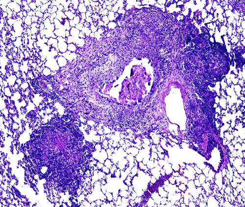
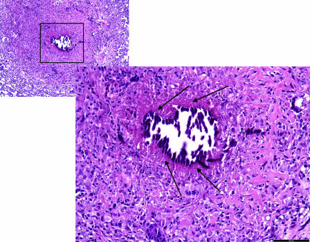
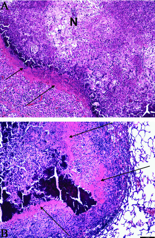
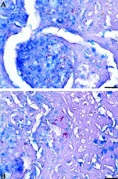
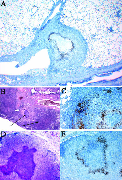
The lengthy chemotherapy of tuberculosis reflects the ability of a small subpopulation of Mycobacterium tuberculosis bacteria to persist in infected individuals. To date, the exact location of these persisting bacteria is not known. Lung lesions in guinea pigs infected with M. tuberculosis have striking similarities, such as necrosis, mineralization, and hypoxia, to natural infections in humans. Guinea pigs develop necrotic primary lesions after aerosol infection that differ in their morphology compared to secondary lesions resulting from hematogenous dissemination. In infected guinea pigs conventional therapy for tuberculosis during 6 weeks reduced the bacterial load by 1.7 logs in the lungs and, although this completely reversed lung inflammation associated with secondary lesions, the primary granulomas remained largely unaffected. Treatment of animals with the experimental drug R207910 (TMC207) for 6 weeks was highly effective with almost complete eradication of the bacteria throughout both the primary and the secondary lesions. Most importantly, the few remnants of acid-fast bacilli remaining after R207910 treatment were to be found extracellular, in a microenvironment of residual primary lesion necrosis with incomplete dystrophic calcification. This zone of the primary granuloma is hypoxic and is morphologically similar to what has been described for human lung lesions. These results show that this acellular rim may, therefore, be a primary location of persisting bacilli withstanding drug treatment.


Figures

FIG. 1.
FIG. 1.
Lungs from an M. tuberculosis-infected guinea pig showing primary and secondary lung lesions. A residual primary lung lesion with dystrophic calcification, which is surrounded by mixed inflammation representing secondary lesions resulting from hematogenous dissemination and inflammation, is shown. Magnification, ×100 (H&E stain).
FIG. 2.
FIG. 2.
Primary granuloma in the lungs of M. tuberculosis-infected guinea pigs after INH-RIF-PZA drug treatment. The image shows a lung from an M. tuberculosis-infected guinea pig treated for 6 weeks with the combination treatment of INH-RIF-PZA; a residual primary lung lesion is evidenced by central necrosis with dystrophic calcification. The lesion is delineated from the normal lung parenchyma by a noncalcified, acellular rim (arrows). The surrounding lung tissue is free of secondary inflammation which is extensive in untreated controls. Magnification ×100 for small inset; magnification ×200 for the large image (H&E stain).
FIG. 3.
FIG. 3.
Detail of the acellular rim delineating the calcified center of the primary granuloma. (A) High magnification of a primary lesion from lungs of an M. tuberculosis-infected guinea pig treated for 6 weeks with the combination therapy INH-RIF-PZA. The residual primary lung lesion is evidenced by central necrosis with incomplete dystrophic calcification (indicated by an “N”). Calcified necrotic debris is the granular basophilic material that forms a well-delineated margin from an acellular, rim (arrows) that blends with the fibrous capsule that contains predominantly lymphocytes and fewer macrophages. The lesion is well delineated from the normal lung parenchyma by a fibrous capsule. Magnification, ×200 (H&E stain). (B) Another high-magnification image of a primary lung lesion from an INH-RIF-PZA treated guinea pig infected with M. tuberculosis. The lesion shows extensive calcification and still has an acellular, rim (arrow) that blends with the fibrous capsule that contains predominantly lymphocytes and fewer macrophages. More normal lung parenchyma is seen at the section margins. Magnification, ×200 (H&E stain).
FIG. 4.
FIG. 4.
Location of AFB in the acellular rim of the primary granuloma. (A and B). Primary lung lesion of an M. tuberculosis-infected guinea pig treated for 6 weeks with R207610; extracellular AFB (stained in red) are in microcolonies primarily within the acellular, uncalcified remnant of necrosis and to a lesser extent within the central area of partially calcified lytic necrosis. (A) The remaining AFB are extracellular in between lymphocytes and macrophages. (B) The remaining AFB are present in a complete acellular region. Magnification, ×400 (acid fast staining).
FIG. 5.
FIG. 5.
Lungs and mesenteric lymph nodes from guinea pigs showing the hypoxic regions in the primary granulomas 4 months after M. tuberculosis infection. (A) Residual primary lung lesion near a major airway with central necrosis surrounded by epithelioid macrophages that are stained by immunohistochemistry for pimonidazole adducts. The center of the lesion, likely hypoxic, fails to stain due to the lack of viable cells. Magnification, ×40 (hematoxylin counterstain). (B) Residual primary lung lesion with dystrophic calcification and secondary lesion resulting from hematogenous dissemination and inflammation Magnification, ×40 (H&E stain), both indicated by arrows. (C) Primary lung lesion with central necrosis surrounded by epithelioid macrophages that are stained by immunohistochemistry for pimonidazole adducts. Magnification, ×100 (hematoxylin counterstain). (D) Mediastinal lymph node where the normal architecture is replaced by mixed inflammation composed primarily of macrophages and with an extensive, central focus of lytic necrosis. Magnification, ×40 (H&E stain). (E) Mediastinal lymph node with the area of central necrosis surrounded by epithelioid macrophages that are stained by immunohistochemistry for pimonidazole adducts. The center of the lesion, likely hypoxic, fails to stain due to the lack of viable cells (necrosis). Magnification, ×40 (hematoxylin counterstain).

References

    1. Aly, S., K. Wagner, C. Keller, S. Malm, A. Malzan, S. Brandau, F. C. Bange, and S. Ehlers. 2006. Oxygen status of lung granulomas in Mycobacterium tuberculosis-infected mice. J. Pathol. 210:298-305. - PubMed
    1. Andries, K., P. Verhasselt, J. Guillemont, H. W. Gohlmann, J. M. Neefs, H. Winkler, J. Van Gestel, P. Timmerman, M. Zhu, E. Lee, P. Williams, D. de Chaffoy, E. Huitric, S. Hoffner, E. Cambau, C. Truffot-Pernot, N. Lounis, and V. Jarlier. 2005. A diarylquinoline drug active on the ATP synthase of Mycobacterium tuberculosis. Science 307:223-227. - PubMed
    1. Arteel, G. E., R. G. Thurman, and J. A. Raleigh. 1998. Reductive metabolism of the hypoxia marker pimonidazole is regulated by oxygen tension independent of the pyridine nucleotide redox state. Eur. J. Biochem. 253:743-750. - PubMed
    1. Arteel, G. E., R. G. Thurman, J. M. Yates, and J. A. Raleigh. 1995. Evidence that hypoxia markers detect oxygen gradients in liver: pimonidazole and retrograde perfusion of rat liver. Br. J. Cancer 72:889-895. - PMC - PubMed
    1. Basaraba, R. J., D. D. Dailey, C. T. McFarland, C. A. Shanley, E. E. Smith, D. N. McMurray, and I. M. Orme. 2006. Lymphadenitis as a major element of disease in the guinea pig model of tuberculosis. Tuberculosis 86:386-394. - PubMed

Publication types

MeSH terms