. 2007 Aug;75(8):3823-32.
doi: 10.1128/IAI.01335-06. Epub 2007 May 21.

Cytokines, signaling pathways, and effector molecules required for the control of Leishmania (Viannia) braziliensis in mice

Affiliations

Cytokines, signaling pathways, and effector molecules required for the control of Leishmania (Viannia) braziliensis in mice

F Janaina Soares Rocha et al. Infect Immun. 2007 Aug.

Abstract

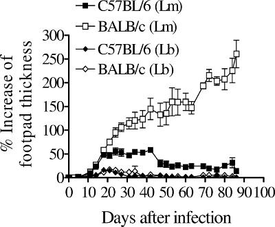
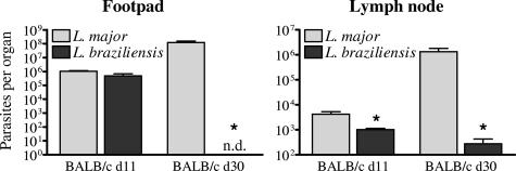
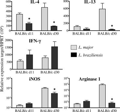
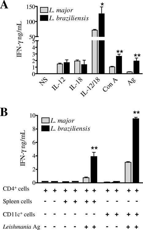
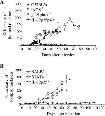
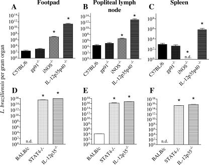
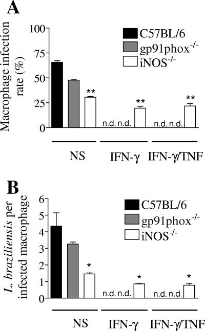
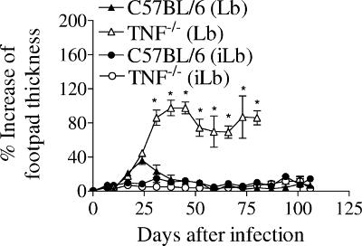
Cutaneous leishmaniasis is caused by protozoan parasites of the genus Leishmania. The mechanisms of pathogen control have been established primarily in the mouse model of Leishmania major infection, but they might not hold true for other Leishmania species associated with cutaneous disease. Here, we analyzed the role of cytokines, signaling components, and effector molecules in the control of New World cutaneous leishmaniasis due to L. braziliensis. Unlike L. major, L. braziliensis caused small, nonulcerative, and self-healing skin swelling in C57BL/6 mice, as well as BALB/c mice. In contrast to the results obtained for L. mexicana, mice deficient for interleukin-12 or its key signaling molecule, signal transducer and activator of transcription 4, rapidly succumbed to severe visceral leishmaniasis. Infection of tumor necrosis factor knockout mice with L. braziliensis led to progressive, nonhealing skin lesions with erosions and hemorrhagic ulcerations, but in contrast to the results with L. major, only 20 to 30% of the mice developed fatal visceral disease. As seen with L. major, mice with a deleted inducible nitric oxide synthase gene (iNOS(-/-)) were unable to contain L. braziliensis in the skin, whereas the control of the parasite in the spleen remained unimpaired. Unlike what happens in L. major infections, NADPH oxidase had no impact on the course of disease in L. braziliensis-infected mice. These results not only define essential components of a protective immune response to L. braziliensis but also illustrate that the requirements for the control of cutaneous leishmaniasis vary between different parasite species.


Figures

FIG. 1.
FIG. 1.
Courses of L. major and L. braziliensis infection in BALB/c and C57BL/6 mice. Groups of four or five BALB/c mice were infected in the right hind footpad with 2 × 10⁶ L. major (Lm) or L. braziliensis (Lb) promastigotes, and the percent increase in footpad thickness (mean ± standard deviation) was determined (see Material and Methods). From day 23 onward, the lesions of L. major-infected BALB/c and C57BL/6 mice were significantly different (P < 0.01 or P < 0.005), whereas the lesions of L. braziliensis-infected BALB/c and C57BL/6 were not significantly different (P > 0.05) (data not shown). The data are representative of three independent experiments in which similar results were obtained.
FIG. 2.
FIG. 2.
Tissue parasite burdens in the skin lesions and draining lymph nodes of BALB/c mice infected with L. major or L. braziliensis. Groups containing eight BALB/c mice each were infected in the right and left hind footpads with 2 × 10⁶ L. major or L. braziliensis promastigotes, and the numbers of viable parasites in the right footpad and draining popliteal lymph node were determined by limiting dilution analysis at day 11 (d11) and day 30 (d30) of infection. The data are the means ± standard errors of the means for the parasite loads of four individually analyzed mice per time point and parasite species. An asterisk indicates that there is a significant difference (P < 0.02) between L. major- and L. braziliensis-infected mice. n.d., no parasites detected. The results of one of two independent experiments are shown.
FIG. 3.
FIG. 3.
Cytokine mRNA expression in the draining lymph nodes of BALB/c mice during the early phase (day 11) and clinically acute phase (day 30) of infection with L. major or L. braziliensis. Groups containing eight BALB/c mice each were infected in the right and left hind footpads with 2 × 10⁶ L. major or L. braziliensis promastigotes. At days 11 (d11) and 30 (d30) after infection total RNA was prepared, and mRNA expression of the cytokines indicated, iNOS, and arginase I was analyzed by real-time RT-PCR. The data are means ± standard errors of the means for the relative gene expression levels in four individually analyzed left lymph nodes per time point and parasite species. An asterisk indicates that there is a significant difference (P < 0.02) between L. major- and L. braziliensis-infected lymph nodes. These data and the data in Fig. 2 were derived from the same infected mice. The results of one of two independent experiments are shown.
FIG. 4.
FIG. 4.
Production of IFN-γ by total lymph node cells (A) or purified CD4+ T cells from pooled lymph nodes (B) of BALB/c mice infected with 2 × 10⁶ L. major or L. braziliensis promastigotes (day 15 of infection) after in vitro restimulation with cytokines (IL-12 and IL-18), a mitogen (concanavalin A [Con A]), or Leishmania antigen (Ag) for 72 h. (A) Total lymph node cells of three mice per group were individually analyzed at day 15 of infection, and IFN-γ (mean ± standard deviation) was measured by ELISA. One asterisk, P < 0.02; two asterisks, P < 0.005. The results of one of two experiments in which similar results were obtained are shown. (B) CD4+ T cells from pooled lymph nodes of three infected BALB/c mice per group were purified, restimulated in triplicate (in the absence or presence of total spleen cells or purified splenic CD11c+ dendritic cells from naïve BALB/c mice as antigen-presenting cells), and the IFN-γ contents (means ± standard deviations) of the culture supernatants were measured by ELISA. Two asterisks, P < 0.005. Data from one of two experiments in which similar results were obtained are shown. NS, medium alone.
FIG. 5.
FIG. 5.
Clinical course of L. braziliensis infection in mice deficient for IL-12, IL-12 plus IL-23, STAT4, or antimicrobial effector mechanisms. C57BL/6 wild-type, IL-12p35/p40−/−, iNOS−/−, and gp91phox−/− mice (A) and BALB/c wild-type, IL-12p35−/−, and STAT4−/− mice (B) were infected in the right hind footpad with 2 × 10⁶ L. braziliensis promastigotes, and the percent increase in footpad thickness (mean ± standard deviation) was determined. An asterisk indicates that mice succumbed due to fatal dissemination of the parasite. From day 25 or 30 (A) or day 38 (B) onward, the lesions of iNOS−/−, IL-12p35/p40−/−, IL-12p35−/−, and STAT4−/− mice were significantly different (P < 0.01 or P < 0.005 [data not shown]) from the lesions of the corresponding wild-type controls. The data are representative of the data from two independent experiments (each with four or five mice per strain) that yielded comparable results.
FIG. 6.
FIG. 6.
Tissue parasite burden in mice deficient for IL-12, IL-12 plus IL-23, STAT4, or antimicrobial effector mechanisms. C57BL/6 wild-type, IL-12p35/p40−/−, iNOS−/−, and gp91phox−/− mice (A to C) and BALB/c wild-type, IL-12p35−/−, and STAT4−/− mice (D to F) were infected in the right hind footpad with 2 × 10⁶ L. braziliensis promastigotes, and the numbers of viable parasites in the footpad (A and D), draining popliteal lymph node (B and E), and spleen (C and F) were determined by limiting dilution analysis at day 36 (A to C) or day 71 (D to F). The error bars indicate the 95% confidence intervals (an asterisk indicates that a value is significantly different from the wild-type control value [P < 0.05 or P < 0.005]). Similar results were obtained in two independent experiments (each with three mice per time point and group). n.d., no parasites detected.
FIG. 7.
FIG. 7.
iNOS protein expression in the popliteal lymph node of a BALB/c wild-type mouse (A) and a STAT4−/− mouse (B) at day 36 after infection with 2 × 10⁶ L. braziliensis promastigotes in the right hind footpad. Anti-iNOS immunohistology determined using immunoperoxidase staining (red) and hematoxylin counterstaining of the nuclei (blue). Magnification, ×200.
FIG. 8.
FIG. 8.
Fate of L. braziliensis in C57BL/6 wild-type, iNOS−/−, and gp91phox−/− macrophages. Thioglycolate-elicited peritoneal macrophages were cultured in medium alone (NS) or in the presence of cytokines (IFN-γ with or without TNF) for 4 h prior to infection with L. braziliensis promastigotes for 16 h. The percentage of infected macrophages (A), the number of intracellular parasites per infected macrophage (B), and the number of intracellular parasites per 100 macrophages in the culture (not shown) were determined 72 h after infection. Asterisks indicate that the results are significantly different from the results for the wild-type controls (one asterisk, P < 0.01; two asterisks, P < 0.001). The data are representative of four independent experiments in which similar results were obtained.
FIG. 9.
FIG. 9.
Clinical course of L. braziliensis infection in wild-type and TNF−/− mice. Groups of five female C57BL/6 wild-type and TNF−/− mice were infected in the right hind footpad with 2 × 10⁶ nonirradiated (i.e., replicating) (Lb) or irradiated (i.e., nonreplicating) (iLb) live L. braziliensis promastigotes, and the percent increase in footpad thickness (mean ± standard deviation) was determined. From day 32 onward, the lesions of TNF−/− mice were significantly different (P < 0.01 or P < 0.005) from the lesions of the corresponding C57BL/6 controls (indicated by the asterisks). The data are representative of three independent experiments in which similar results were obtained.

Similar articles

Cited by

References

    1. Aguliar Torrentera, F., J. D. Laman, M. V. Meurs, L. Adorini, E. Muraille, and Y. Carlier. 2002. Endogenous interleukin-12 is critical for controlling the late, but not the early stage of Leishmania mexicana infection in C57BL/6 mice. Infect. Immun. 70:5075-5080. - PMC - PubMed
    1. Amato, V. S., H. F. Andrade, V. A. Neto, and M. I. S. Duarte. 2003. Persistence of tumor necrosis factor-α in situ after lesion healing in mucosal leishmaniasis. Am. J. Trop. Med. Hyg. 68:527-528. - PubMed
    1. Anderson, C. F., S. Mendez, and D. L. Sacks. 2005. Nonhealing infection despite Th1 polarization produced by a strain of Leishmania major in C57BL/6 mice. J. Immunol. 174:2934-2941. - PubMed
    1. Barral, A., J. Guerreiro, G. Bomfim, D. Correia, M. Barral-Netto, and E. M. Carvalho. 1995. Lymphadenopathy as the first sign of human cutaneous infection by Leishmania braziliensis. Am. J. Trop. Med. Hyg. 53:256-259. - PubMed
    1. Belosevic, M., D. S. Finbloom, P. H. van der Meide, M. V. Slayter, and C. A. Nacy. 1989. Administration of monoclonal anti-IFN-γ antibodies in vivo abrogates natural resistance of C3H/HeN mice to infection with Leishmania major. J. Immunol. 143:266-274. - PubMed

Publication types

MeSH terms

LinkOut - more resources