. 2007 Aug;75(8):3758-68.
doi: 10.1128/IAI.00225-07. Epub 2007 May 21.

Plasmodium yoelii sporozoites with simultaneous deletion of P52 and P36 are completely attenuated and confer sterile immunity against infection

Affiliations

Plasmodium yoelii sporozoites with simultaneous deletion of P52 and P36 are completely attenuated and confer sterile immunity against infection

Mehdi Labaied et al. Infect Immun. 2007 Aug.

Abstract

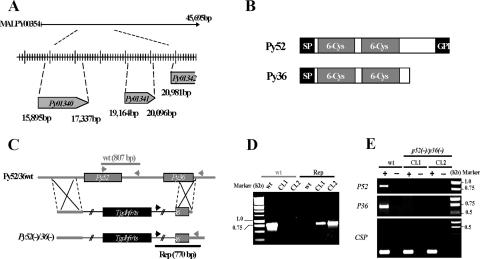
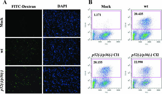
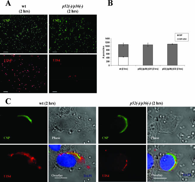
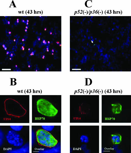
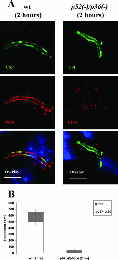
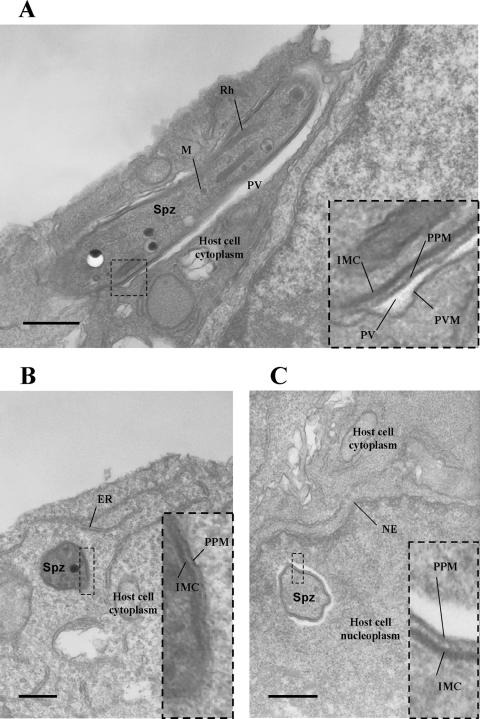
Malaria infection starts when sporozoites are transmitted to the mammalian host during a mosquito bite. Sporozoites enter the blood circulation, reach the liver, and infect hepatocytes. The formation of a parasitophorous vacuole (PV) establishes their intracellular niche. Recently, two members of the 6-Cys domain protein family, P52 and P36, were each shown to play an important albeit nonessential role in Plasmodium berghei sporozoite infectivity for the rodent host. Here, we generated p52/p36-deficient Plasmodium yoelii parasites by the simultaneous deletion of both genes using a single genetic manipulation. p52/p36-deficient parasites exhibited normal progression through the life cycle during blood-stage infection, transmission to mosquitoes, mosquito-stage development, and sporozoite infection of the salivary glands. p52/p36-deficient sporozoites also showed normal motility and cell traversal activity. However, immunofluorescence analysis and electron microscopic observations revealed that p52/p36-deficient parasites did not form a PV within hepatocytes in vitro and in vivo. The p52/p36-deficient parasites localized as free entities in the host cell cytoplasm or the host cell nucleoplasm and did not develop as liver stages. Consequently, they did not cause blood-stage infections even at high sporozoite inoculation doses. Mice immunized with p52/p36-deficient sporozoites were completely protected against infectious sporozoite challenge. Our results demonstrate for the first time the generation of two-locus gene deletion-attenuated parasites that infect the liver but do not progress to blood-stage infection. The study will critically guide the design of Plasmodium falciparum live attenuated malaria vaccines.


Figures

FIG. 1.
FIG. 1.
Targeted gene disruption of P52 and P36 using a single-replacement strategy. (A) P. yoelii P52 (PY01340) and P36 (PY01341) are located in tandem on contig MALPY00354. (B) Predicted protein structure of P52 and P36. Each protein exhibits a signal peptide (SP) followed by two 6-Cys domains. In addition, P52 possesses a putative GPI anchor transfer peptide. (C) Schematic representation of the replacement strategy to generate the p52/p36-deficient parasites. The wild-type (wt) P52/P36 genomic locus was targeted with a replacement plasmid containing a 5′ untranslated region fragment of P52 and a 3′ fragment of the P36 ORF that flank the Toxoplasma gondii dihydrofolate reductase-thymidylate synthase-positive selectable marker. A recombination event (double crossover) resulted in the replacement of the P52 ORF and the 5′ part of the P36 ORF by the selection marker. wt and replacement-specific oligonucleotide primer combinations used for genotyping are indicated by arrows, and expected fragments are shown by gray and black lines. (D) PCR genotyping. Amplification with oligonucleotide primer combinations that can amplify only from the recombinant locus (Rep) confirmed the gene replacement. The wt-specific oligonucleotide primer combinations confirmed the absence of residual wt parasites in p52/p36-deficient Cl1 and Cl2. (E) The p52/p36-deficient parasites do not transcribe P52 and P36. The absence of P52 and P36 transcripts in p52/p36-deficient parasites was shown by RT-PCR using gene-specific oligonucleotide primers and salivary gland sporozoite RNA as a template. Gene-specific oligonucleotide primers for CSP were used as a positive control and amplified from wt and p52/p36-deficient sporozoite RNA. − indicates reactions without RT, and + indicates reactions with RT.
FIG. 2.
FIG. 2.
p52/p36-deficient sporozoites traverse and wound cells normally. (A) Overview of infected HepG2-CD81 cultures showing FITC-positive (green) wounded cells as a result of wt or p52/p36-deficient sporozoite traversal. DAPI (blue) visualizes the nuclei. (B) Quantification of cell-wounding activity using flow cytometry reveals a similar level of traversal activity in wt and p52/p36-deficient sporozoites. The assay was repeated six times for each sample. Approximately 3.0 × 10⁴ sporozoites were incubated with 5.0 × 10⁴ subconfluent HepG2-CD81 cells per well in the presence of FITC-dextran. The x axis represents the forward-scatter properties of the cells, while the y axis represents the green fluorescence. The numbers of wounded cells (percent) are shown in the upper left corners of the graphs. Approximately 20% of the HepG2-CD81 cells inoculated with wt and p52/p36-deficient (Cl1 and Cl2) sporozoites were fluorescent, i.e., wounded. Mock infections were done by incubating HepG2-CD81 cells with uninfected mosquito salivary gland preparations.
FIG. 3.
FIG. 3.
p52/p36-deficient parasites fail to infect hepatocytes with the formation of a PV in vitro. Immunofluorescence assay with infected HepG2-CD81 cells using Abs to UIS4, a PVM-resident protein, allows the detection of the parasite PVM 2 h p.i. (A) Low-magnification images showing wt parasite staining with anti-UIS4 and anti-CSP Abs (left panels). UIS4 expression is not apparent in p52/p36-deficient parasites at the low magnification shown in the right panels (scale bar, 40 μm). (B) Microscopic quantification reveals that ∼40% of wt parasites show strong UIS4 staining of the PVM and show CSP staining of the parasite surface. The remaining ∼60% of wt parasites stained with CSP represent extracellular sporozoites and sporozoites in cell traversal mode. p52/p36-deficient parasites were detected by anti-CSP staining only. Numbers shown are means ± standard deviations of counting 1.0 × 10³ parasites per well in three wells. (C) Higher magnification shows typical peripheral localization of UIS4 in the PVM surrounding an intracellular wt parasite. Intracellular p52/p36-deficient parasites occasionally exhibit a signal of UIS4 staining within the sporozoite (scale bar, 10 μm). Cells were labeled with DAPI (blue) to visualize the nuclei.
FIG. 4.
FIG. 4.
p52/p36-deficient parasites show a severe defect in LS development in vitro. P. yoelii wt LSs grow in HepG2-CD81 cells and complete development, but p52/p36-deficient LSs do not grow and do not persist in infected cells. (A) Forty-three hours p.i., wt LSs have developed to the late schizont stage. LSs are visualized by staining with anti-UIS4 Abs (red) and anti-Hsp70 Abs (green). Nuclei were visualized with DAPI (scale bar, 30 μm). (B) Higher magnification shows a late wt LS with multiple nuclei entirely surrounded by a PVM that is revealed by anti-UIS4 staining (scale bar, 5 μm). (C) p52/p36-deficient parasites do not grow in HepG2-CD81 cells. A small p52/p36-deficient growth-arrested LS is indicated by the white arrow (scale bar, 30 μm). (D) Higher magnification of a growth-arrested p52/p36-deficient parasite 43 h p.i. does not show a typical PVM using UIS4 staining. Some UIS4-positive vesicular structures are visible (scale bar, 5 μm).
FIG. 5.
FIG. 5.
p52/p36-deficient parasites fail to establish infection in the host liver. (A) Indirect immunofluorescence assay of wt and p52/p36-deficient parasites detected in the host liver at 2 h p.i. The upper panels show intracellular wt parasite or p52/p36-deficient parasite staining with anti-CSP Abs. The PVM is visualized in wt parasites using anti-UIS4 Ab staining, but UIS4 staining is not discernable in p52/p36-deficient parasites, indicating a PVM deficiency (middle panels). The overlay of UIS4 staining, CSP staining, and nuclear DAPI staining is shown in the bottom panels (scale bar, 20 μm). (B) Quantification of liver infections. Numbers shown are means ± standard deviations of wt parasites and p52/p36-deficient parasites detected in three discontinuous sections of livers of BALB/c mice 2 h p.i. p52/p36-deficient parasites were detected at greatly reduced numbers (∼90% reduction) compared to wt parasites and did not show UIS4 staining (P < 0.0001, Fisher's exact test). Approximately 75% of wt parasites detected at 2 h p.i. showed UIS4-positive staining and CSP-positive staining.
FIG. 6.
FIG. 6.
Electron microscopic analysis confirms that p52/p36-deficient parasites cannot form a parasitophorous vacuole. (A) wt sporozoite (longitudinal view) within a HepG2-CD81 cell 1 h after infection. The parasite is surrounded by a PVM. (B) p52/p36-deficient sporozoite (transversal view) within a HepG2-CD81 cell 1 h after infection. The parasite lacks a PVM and appears to be in direct contact with the host cell cytoplasm. (C) A p52/p36-deficient sporozoite (transversal view) was also detected within the host cell nucleus, surrounded by nucleoplasm. All scale bars are 0.5 μm. The inset boxes show higher magnifications of the boxed areas within the overview images. ER, endoplasmic reticulum; IMC, inner membrane complex; M, microneme; NE, nuclear envelope; PPM, parasite plasma membrane; Spz, sporozoite; Rh, rhoptry.

Similar articles

Cited by

References

    1. Baer, K., M. Roosevelt, A. B. Clarkson, Jr., N. van Rooijen, T. Schnieder, and U. Frevert. 2007. Kupffer cells are obligatory for Plasmodium yoelii sporozoite infection of the liver. Cell. Microbiol. 9:397-412. - PubMed
    1. Belmonte, M., T. R. Jones, M. Lu, R. Arcilla, T. Smalls, A. Belmonte, J. Rosenbloom, D. J. Carucci, and M. Sedegah. 2003. The infectivity of Plasmodium yoelii in different strains of mice. J. Parasitol. 89:602-603. - PubMed
    1. Carter, R., A. Coulson, S. Bhatti, B. J. Taylor, and J. F. Elliott. 1995. Predicted disulfide-bonded structures for three uniquely related proteins of Plasmodium falciparum, Pfs230, Pfs48/45 and Pf12. Mol. Biochem. Parasitol. 71:203-210. - PubMed
    1. Eksi, S., B. Czesny, G. J. van Gemert, R. W. Sauerwein, W. Eling, and K. C. Williamson. 2006. Malaria transmission-blocking antigen, Pfs230, mediates human red blood cell binding to exflagellating male parasites and oocyst production. Mol. Microbiol. 61:991-998. - PubMed
    1. Frevert, U., S. Engelmann, S. Zougbede, J. Stange, B. Ng, K. Matuschewski, L. Liebes, and H. Yee. 2005. Intravital observation of Plasmodium berghei sporozoite infection of the liver. PLoS Biol. 3:e192. - PMC - PubMed

Publication types

MeSH terms

Substances