. 2007 Aug;75(8):3979-88.
doi: 10.1128/IAI.00290-07. Epub 2007 May 21.

Mycolactone-mediated inhibition of tumor necrosis factor production by macrophages infected with Mycobacterium ulcerans has implications for the control of infection

Affiliations

Mycolactone-mediated inhibition of tumor necrosis factor production by macrophages infected with Mycobacterium ulcerans has implications for the control of infection

Egídio Torrado et al. Infect Immun. 2007 Aug.

Abstract

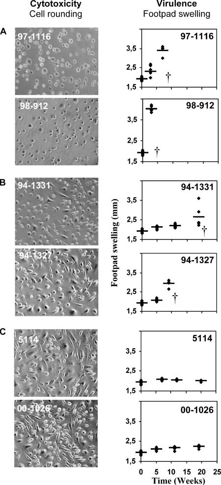
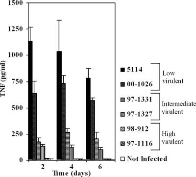
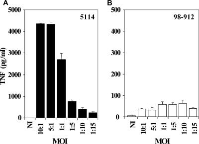
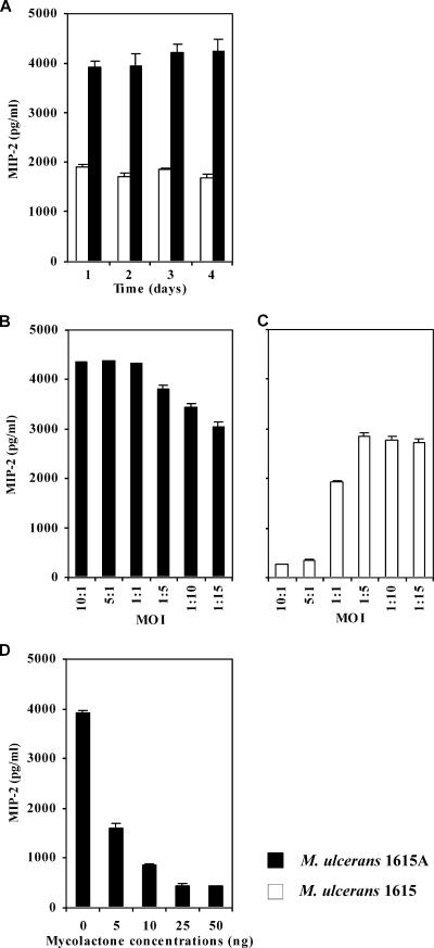
The pathogenicity of Mycobacterium ulcerans, the agent of Buruli ulcer, depends on the cytotoxic exotoxin mycolactone. Little is known about the immune response to this pathogen. Following the demonstration of an intracellular growth phase in the life cycle of M. ulcerans, we investigated the production of tumor necrosis factor (TNF) induced by intramacrophage bacilli of diverse toxigenesis/virulence, as well as the biological relevance of TNF during M. ulcerans experimental infections. Our data show that murine bone marrow-derived macrophages infected with mycolactone-negative strains of M. ulcerans (nonvirulent) produce high amounts of TNF, while macrophages infected with mycolactone-positive strains of intermediate or high virulence produce intermediate or low amounts of TNF, respectively. These results are in accordance with the finding that TNF receptor P55-deficient (TNF-P55 KO) mice are not more susceptible than wild-type mice to infection by the highly virulent strains but are more susceptible to nonvirulent and intermediately virulent strains, demonstrating that TNF is required to control the proliferation of these strains in animals experimentally infected by M. ulcerans. We also show that mycolactone produced by intramacrophage M. ulcerans bacilli inhibits, in a dose-dependent manner, but does not abrogate, the production of macrophage inflammatory protein 2, which is consistent with the persistent inflammatory responses observed in experimentally infected mice.


Figures

FIG. 1.
FIG. 1.
Cytotoxic activity and virulence of different strains of M. ulcerans. (Left) BMDM were infected with different strains of M. ulcerans at an MOI (bacilli/macrophages) of 1:1. Macrophages were photographed by phase-contrast microscopy 6 days after infection. M. ulcerans cytotoxicity was determined based on the microscopic observation of cell rounding and detachment from the monolayer. (Right) Mice were infected in the left hind footpad with 5.3 log10 AFB of different M. ulcerans strains. Virulence was determined by measuring footpad swelling. For ethical reasons mice were sacrificed after the emergence of ulceration. Results are from one representative experiment out of three independent experiments.
FIG. 2.
FIG. 2.
TNF production by BMDM infected with strains of M. ulcerans that produce different amounts and types of mycolactone. Strains of M. ulcerans from different origins were used to infect BMDM at an MOI of 1:1. At days 2, 4, and 6 postinfection the supernatants of three independent wells were removed for each strain and frozen until cytokine measurement by ELISA. Low amounts of TNF were produced by macrophages infected with high cytotoxic/virulent strains (98-912 and 97-1116), whereas high amounts of TNF were produced in the case of the low cytotoxicity/low virulence strain 5114 or M. marinum 00-1026. Intermediate levels of TNF are produced with strains of intermediate cytotoxicity/virulence (94-1327 and 94-1331). Error bars indicate standard deviations. Results are from one representative experiment out of three independent experiments.
FIG. 3.
FIG. 3.
TNF production by BMDM infected at different MOIs with the mycolactone A/B-producing strain 98-912 or the mycolactone-negative strain 5114. BMDM were infected at MOIs ranging from 10:1 to 1:15 with the M. ulcerans strains 5114 (A) or 98-912 (B). Twenty-four hours postinfection, the supernatant from three independent wells was removed for each strain and stored frozen until cytokine measurement by ELISA. BMDM infected with M. ulcerans 5114 produce high amounts of TNF at high MOIs, and TNF production decreases as the MOI decreases. With M. ulcerans 98-912, production of TNF is low, independent of the MOI. Results are from one representative experiment out of three independent experiments.
FIG. 4.
FIG. 4.
TNF production by BMDM infected with WT M. ulcerans 1615 or with the mycolactone-defective mutant 1615A: kinetics and effects of MOI and supplementation with mycolactone. BMDM were infected with a 1:1 MOI (A and D) or at different MOIs ranging from 10:1 to 1:15 (B and C) with M. ulcerans 1615A or 1615 WT, in the presence (D) or absence (A to C) of mycolactone. For cytokine measurement by ELISA, supernatants of three independent wells were collected from each experimental group. (A) Higher amounts of TNF are produced with infection by M. ulcerans 1615A in comparison to M. ulcerans 1615. (B and C) BMDM infected with M. ulcerans 1615A produce large amounts of TNF at high MOIs, and that production declines as the MOI is decreased. M. ulcerans 1615 induces the production of smaller amounts of TNF peaking at low MOI. (D) Addition of mycolactone to M. ulcerans 1615A-infected macrophages diminishes or abrogates the production of TNF. TNF was not detected in the presence of 50 ng mycolactone. Results are from one representative experiment out of two independent experiments.
FIG. 5.
FIG. 5.
MIP-2 production by BMDM infected with WT M. ulcerans 1615 or with the mycolactone-defective mutant 1615A: kinetics and effects of MOI and supplementation with mycolactone. BMDM were infected with a 1:1 MOI (A and D) or with MOIs ranging from 10:1 to 1:15 (B and C) of M. ulcerans strain 1615A or 1615, in the presence (D) or absence (A to C) of mycolactone. For cytokine measurement by ELISA, supernatants of three independent wells were collected from each experimental group. (A) Although M. ulcerans 1615A induces larger amounts of MIP-2 in comparison with M. ulcerans 1615, significant amounts of the cytokine are produced with the mycolactone-positive strain. (B and C) BMDM infected with M. ulcerans 1615A produce large amounts of MIP-2 irrespective of MOI. M. ulcerans 1615 induces the production of small but important amounts of MIP-2 at low MOIs. (D) Addition of mycolactone to M. ulcerans 1615A-infected macrophages diminishes but does not abrogate the production of MIP-2, even at high concentrations. Results are from one representative experiment out of two independent experiments.
FIG. 6.
FIG. 6.
Proliferation of the mycolactone A/B-producing strain 98-912, mycolactone C-producing strain 94-1327, or mycolactone-negative strain 5114 in wild-type and TNF receptor-deficient mice. Mice (WT [closed symbols] or TNF-P55 KO [open symbols]) were infected subcutaneously in the left hind footpad with 5.3 log10 AFB of M. ulcerans 5114 (diamonds), 94-1327 (triangles), or 98-912 (squares). The number of AFB in homogenates from four footpads in each infected group was counted at the indicated time points. WT mice control the proliferation of M. ulcerans 5114 but do not eliminate the infection, whereas TNF-P55 KO mice are more susceptible to this strain. An increase in bacterial counts was also found in the footpads of TNF-P55 KO mice infected with strain 94-1327 compared with the WT mice. No significant differences were found for mice infected with M. ulcerans 98-912. For ethical reasons, mice were sacrificed after the emergence of ulceration. Statistical differences were determined by comparing TNF-P55 KO mice with WT mice. Calculations were performed using Student's t test. *, P < 0.05; **, P < 0.01; ***, P < 0.001. Results are from one representative experiment out of two independent experiments.

Similar articles

Cited by

References

    1. Abalos, F. M., J. Aguiar, Sr., A. Guedenon, F. Portaels, and W. M. Meyers. 2000. Mycobacterium ulcerans infection (Buruli ulcer): a case report of the disseminated nonulcerative form. Ann. Diagn. Pathol. 4:386-390. - PubMed
    1. Adusumilli, S., A. Mve-Obiang, T. Sparer, W. Meyers, J. Hayman, and P. L. Small. 2005. Mycobacterium ulcerans toxic macrolide, mycolactone modulates the host immune response and cellular location of M. ulcerans in vitro and in vivo. Cell. Microbiol. 7:1295-1304. - PubMed
    1. Asiedu, K., and S. Etuaful. 1998. Socioeconomic implications of Buruli ulcer in Ghana: a three-year review. Am. J. Trop. Med. Hyg. 59:1015-1022. - PubMed
    1. Bean, A. G., D. R. Roach, H. Briscoe, M. P. France, H. Korner, J. D. Sedgwick, and W. J. Britton. 1999. Structural deficiencies in granuloma formation in TNF gene-targeted mice underlie the heightened susceptibility to aerosol Mycobacterium tuberculosis infection, which is not compensated for by lymphotoxin. J. Immunol. 162:3504-3511. - PubMed
    1. Bendtzen, K. 1988. Interleukin 1, interleukin 6 and tumor necrosis factor in infection, inflammation and immunity. Immunol. Lett. 19:183-191. - PubMed

Publication types

MeSH terms