. 2007 May 21;177(4):579-85.
doi: 10.1083/jcb.200611046.

Chemoattractants and chemorepellents act by inducing opposite polarity in phospholipase C and PI3-kinase signaling

Affiliations

Chemoattractants and chemorepellents act by inducing opposite polarity in phospholipase C and PI3-kinase signaling

Ineke Keizer-Gunnink et al. J Cell Biol. .

Abstract

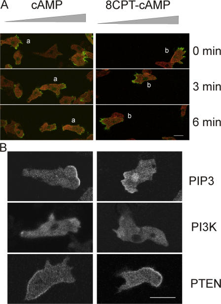
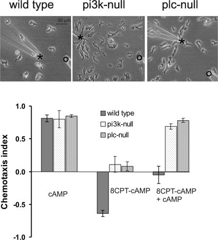
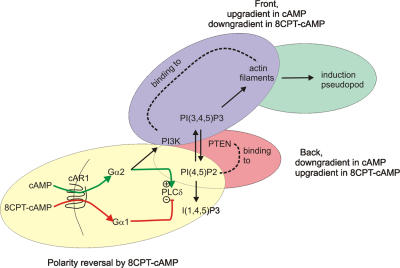
During embryonic development, cell movement is orchestrated by a multitude of attractants and repellents. Chemoattractants applied as a gradient, such as cAMP with Dictyostelium discoideum or fMLP with neutrophils, induce the activation of phospholipase C (PLC) and phosphoinositide 3 (PI3)-kinase at the front of the cell, leading to the localized depletion of phosphatidylinositol 4,5-bisphosphate (PI[4,5]P(2)) and the accumulation of phosphatidylinositol-3,4,5-trisphosphate (PI[3,4,5]P(3)). Using D. discoideum, we show that chemorepellent cAMP analogues induce localized inhibition of PLC, thereby reversing the polarity of PI(4,5)P(2). This leads to the accumulation of PI(3,4,5)P(3) at the rear of the cell, and chemotaxis occurs away from the source. We conclude that a PLC polarity switch controls the response to attractants and repellents.


Figures

Figure 1.
Figure 1.
Antagonism of chemotaxis to cAMP by 8CPT-cAMP. Starved D. discoideum cells were spread on a polystyrene surface in a droplet with 0.5 ml of 10 mM phosphate buffer, pH 6.1, at room temperature, and stimulated by micropipettes filled with 10 mM 8CPT-cAMP (bottom right, open circle) or 0.1 mM cAMP (top left, asterisk); see Materials and methods for further details. (A) Four frames from a movie (Video 3) showing the distribution of cells without stimulus, 8 min after stimulation with 8CPT-cAMP, 5 min after stimulation with 8CPT-cAMP and cAMP, and 12 min after stimulation with cAMP alone (by removing the pipette with 8CPT-cAMP). (B) The chemotaxis index was determined for 20 cells that were shown in Video 3; (top) the filled sections show the simulation with cAMP and/or 8CPT-cAMP. (C) The chemotaxis index was calculated for 85 cells from 6 independent experiments; data shown are the mean ± the SEM. Video 3 is available at http://www.jcb.org/cgi/content/full/jcb.200611046/DC1.
Figure 2.
Figure 2.
Confocal fluorescent images of cells stimulated with cAMP or 8CPT-cAMP. (A) Cells expressing myosin II-RFP (red) and the F-actin–binding protein LimE-GFP (green) were stimulated with cAMP or 8CPT-cAMP by pipettes that are positioned at the right. The figure shows 3 frames of a movie with 5-min intervals (cAMP, Video 4; 8CPT-cAMP, Video 5). (B) Cells expressing the PI(3,4,5)P3 detector PHcrac-GFP, PI3K-GFP, or PTEN-GFP were stimulated with cAMP or 8CPT-cAMP by pipettes that are positioned at the right. The figure shows a representative cell for each case (a field of cells expressing PHcrac-GFP is presented in Video 4 for cAMP and Videos 5 and 6 for 8CPT-cAMP). Videos 4–6 are available at http://www.jcb.org/cgi/content/full/jcb.200611046/DC1.
Figure 3.
Figure 3.
Chemotaxis of mutants with deletions of PI3K or PLC. (A) Frames from movies presenting the distribution of cells at 15 min after stimulation with both 8CPT-cAMP (open circles) and cAMP (asterisks) taken from Video 3 for WT cells, Video 8 for pi3k1/2-null cells, and Video 9 for plc-null cells. Wild-type cells show random distribution, whereas pi3k1/2-null and plc-null cells are attracted toward the pipette with cAMP. (B) Chemotaxis index of wild-type, pi3k1/2-null, and plc-null cells toward cAMP, 8CPT-cAMP, or 8CPT-cAMP and cAMP. The results show that wild-type cells move away from 8CPT-cAMP, whereas pi3k1/2-null and plc-null are not repelled from 8CPT-cAMP. Moreover, chemotaxis toward cAMP is antagonized by 8CPT-cAMP in wild-type cells, but not in pi3k1/2-null and plc-null cells. Videos 3, 8, and 9 are available at http://www.jcb.org/cgi/content/full/jcb.200611046/DC1.
Figure 4.
Figure 4.
Model for polarity reversal leading to cAMP-induced attraction and 8CPT-cAMP–induced repulsion. The model contains three regulatory loops: first, a PLC activation/inhibition loop providing primary polarity of the PI(4,5)P2 gradient; second, a PI(4,5)P2/PTEN loop providing degradation of PI(3,4,5)P3; and third, a PI3K/F-actin loop providing PI(3,4,5)P3-mediated pseudopod extension. Because of inhibition of PLC by 8CPT-cAMP compared with stimulation by cAMP, the PI(4,5)P2 polarity inverses, PI(3,4,5)P3 accumulates downgradient, and cells move away from the pipette. The scheme is based on the observation that the PI(3,4,5)P3-degrading enzyme PTEN binds to PI(4,5)P2, whereas PI3K binds to actin filaments in the leading edge (Iijima et al., 2004; Sasaki et al., 2004), and on the observation that cAMP activates PLC, whereas 8CPT-cAMP inhibits this enzyme (Bominaar and Van Haastert, 1993, 1994; Peters et al., 1991). The mediating G proteins have been identified using knock-out cells (see supporting biochemical data in Fig. S1). Fig. S1 is available at http://www.jcb.org/cgi/content/full/jcb.200611046/DC1.

References

    1. Affolter, M., and C.J. Weijer. 2005. Signaling to cytoskeletal dynamics during chemotaxis. Dev. Cell. 9:19–34. - PubMed
    1. Baggiolini, M. 1998. Chemokines and leukocyte traffic. Nature. 392:565–568. - PubMed
    1. Bominaar, A.A., F. Kesbeke, and P.J.M. Van Haastert. 1994. Phospholipase C in Dictyostelium discoideum. Cyclic AMP surface receptor and G-protein-regulated activity in vitro. Biochem. J. 297:181–187. - PMC - PubMed
    1. Bominaar, A.A., and P.J.M. Van Haastert. 1993. Chemotactic antagonists of cAMP inhibit Dictyostelium phospholipase C. J. Cell Sci. 104:181–185. - PubMed
    1. Bominaar, A.A., and P.J.M. Van Haastert. 1994. Phospholipase C in Dictyostelium discoideum. Identification of stimulatory and inhibitory surface receptors and G-proteins. Biochem. J. 297:189–193. - PMC - PubMed

MeSH terms

Substances