. 2007 May 21;177(4):625-36.
doi: 10.1083/jcb.200610148.

BimS-induced apoptosis requires mitochondrial localization but not interaction with anti-apoptotic Bcl-2 proteins

Affiliations

BimS-induced apoptosis requires mitochondrial localization but not interaction with anti-apoptotic Bcl-2 proteins

Arnim Weber et al. J Cell Biol. .

Abstract

Release of apoptogenic proteins such as cytochrome c from mitochondria is regulated by pro- and anti-apoptotic Bcl-2 family proteins, with pro-apoptotic BH3-only proteins activating Bax and Bak. Current models assume that apoptosis induction occurs via the binding and inactivation of anti-apoptotic Bcl-2 proteins by BH3-only proteins or by direct binding to Bax. Here, we analyze apoptosis induction by the BH3-only protein Bim(S). Regulated expression of Bim(S) in epithelial cells was followed by its rapid mitochondrial translocation and mitochondrial membrane insertion in the absence of detectable binding to anti-apoptotic Bcl-2 proteins. This caused mitochondrial recruitment and activation of Bax and apoptosis. Mutational analysis of Bim(S) showed that mitochondrial targeting, but not binding to Bcl-2 or Mcl-1, was required for apoptosis induction. In yeast, Bim(S) enhanced the killing activity of Bax in the absence of anti-apoptotic Bcl-2 proteins. Thus, cell death induction by a BH3-only protein can occur through a process that is independent of anti-apoptotic Bcl-2 proteins but requires mitochondrial targeting.


Figures

Figure 1.
Figure 1.
Interaction between BimS and prosurvival proteins as tested by immunoprecipitation. BimS-expressing clone A2 (a, c, and d) or T-Rex-293 cells transiently transfected with the BimS construct (b) were left untreated or treated with tetracycline for 6 h (A2 clone) or 16 h (T-Rex-293 cells). Mitochondrial lysates (a–c) or whole-cell lysates (d) were immunoprecipitated with anti-Bim antibodies. Aliquots of mitochondrial (L, 45 μg of protein) or total cell lysates (L, 22 μg), post-IP supernatant (SN), and IP pellets (IP) were loaded and membranes were probed for Bim, Bcl-2, Mcl-1, Bcl-w, A1, and Bcl-xL. The migration of immunoglobulin light chain (Ig) is indicated but was not observed in all experiments. (c) Left, pellet fractions after Bim-IP from isolated mitochondria (first lane), cytosolic fraction (middle lane) after 4 h of tet and from mitochondria without tet (third lane). Right, specificity control with antibodies against an unrelated protein (mBmf). (d) Binding of tet-induced BimS to transiently expressed Bcl-2. Cells were transfected with control vector or an expression vector for human Bcl-2, lysed, and Bim was immunoprecipitated as above. Bim and Bcl-2 were detected by Western blotting. All Western blots are representative of three independent experiments (the experiment shown in panel d was done twice).
Figure 2.
Figure 2.
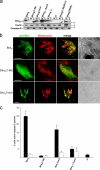
BimS localizes to and inserts into mitochondrial membranes in HeLa cells and in isolated mitochondria. (a) Mitochondria (m) from T-REx-HeLa cells (A2 clone) uninduced or tet-induced as indicated were isolated and separated alongside the soluble cytosolic fractions (c) on 12.5% SDS-PAA gels. CoxIV is a marker for mitochondria, caspase-8 for cytosolic proteins. (b) Isolated mitochondria were treated with 0.1 M Na2CO3 to separate membrane inserted (Ins.) from soluble (Att., membrane attached, intermembrane space and matrix) fractions. Both fractions were analyzed by Western blotting for Bim, CoxIV as a marker for mitochondrial membranes and Bax. (c) Import of in vitro–translated BimEL or BimS into isolated HeLa mitochondria. Radiolabeled precursors of BimEL and BimS were incubated for 10 and 30 min at 30°C with mitochondria isolated from HeLa cells. Subsequently, the samples were split. One third was treated with 50 μg/ml proteinase K (PK) for 20 min on ice, another one was left untreated. The last third was subjected to alkaline extraction resulting in a pellet fraction (P; containing integral membrane proteins) and a supernatant fraction (S; containing soluble and peripherally attached proteins). For comparison, 10% of the total input of radiolabeled precursors was included (T). Mitochondria were reisolated and import was analyzed by SDS-PAGE and autoradiography. *, very likely proteolysis products.
Figure 3.
Figure 3.
BimS-expression causes Bax oligomerization and mitochondrial recruitment of Bax. (a) BimS was induced in HeLa cells (A2 clone) as indicated for 6 h. Mitochondria were isolated and treated with two different cross-linkers as described in Materials and Methods. After cross-linking, the reaction was analyzed by Western blotting with an anti-Bax monoclonal antibody. (b) BimS cells (A2 clone) were transiently transfected with the EGFP-Bax fusion construct and were analyzed by confocal microscopy untreated (top) or upon BimS-induction (bottom, 5 h). Localization of EGFP-Bax (green), mitochondria (red), BimS (blue), and various overlays are shown. Bars, 10 μm.
Figure 4.
Figure 4.
Schematic representation of BimS and BimS mutant proteins used in this study. Mutants were generated that lack the hydrophobic C terminus (1–88, bottom) or where the C terminus was replaced with the mitochondrial membrane anchor of yeast Tom5 (BimSTom5, middle) or that contain various mutations in the BH3 domain as indicated in the box.
Figure 5.
Figure 5.
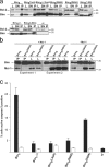
Subcellular localization of BimS mutants and their pro-apoptotic activity. (a) Localization as determined by cell fractionation. T-REx-293 cells were transiently transfected with wild-type or mutant constructs and induced 7 h later with tet for ∼15 h. Mitochondria (m) and cytosolic fractions (c) were prepared and tested for expression of BimS proteins. As a control (ct) the cells were transfected with the empty expression vector. BimS(1–88)/Bcl-2: cells were cotransfected with BimS(1–88) and an expression construct for hBcl-2. The mutants are described in Fig. 4. (b) Localization of C-terminal mutants as determined by confocal microscopy. Cells were transfected as above, induced 24 h later for 5 h with tet, and BimS was detected by staining with Bim-specific antibodies. Mitochondria were identified by MitoTracker staining. Bars, 10 μm. (c) T-Rex-293 cells were transiently transfected with the constructs indicated (ct, empty vector control). 7 h later, expression of BimS was induced by tet addition as shown. 15 h later, cells were harvested and percentage of active caspase-3–positive cells was determined by flow cytometry. Black bars indicate induction with tet; white bars without tet. The values give the mean of at least three independent experiments/SEM. Expression of transfected proteins was confirmed by Western blotting (not depicted).
Figure 6.
Figure 6.
Bcl-2/Mcl-1 binding and pro-apoptotic activity of BH3 domain mutants of BimS. T-REx-293 cells were transfected with the mutants indicated (see Fig. 4) together with an expression construct for hBcl-2 (a) or alone (c). After 7 h, BimS was induced with tet. (a) 18 h later Bim was immunoprecipitated from whole-cell extracts (200 μg) of T-REx-293 cells as above. Control (ct), cells were transfected with hBcl-2 vector and the empty expression vector instead of a BimS expression vector. (b) T-REx-293 cells were cotransfected with hMcl-1 vector and the wild-type BimS or the mutant construct BimS(D69A). 18 h later Bim was immunoprecipitated from whole-cell extracts (200 μg left, 300 μg middle and right) as above. Two experiments are shown. Right: same experiment showing binding of endogenous Mcl-1 to Bim where cells were solely transfected with wt BimS or the mutant construct BimS(D69A). (c) 15 h later cells positive for active caspase-3 were scored by flow cytometry after staining with a monoclonal antibody against active caspase-3 and a Cy3-conjugated secondary antibody. Values give mean of at least three independent experiments/SEM. Expression of transfected proteins was confirmed by Western blotting (not depicted).
Figure 7.
Figure 7.
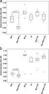
BimS accelerates the growth inhibiting effect of Bax in yeast. Yeast strains containing BimS, Bax, or Bax/BimS were grown in the presence or absence of tetracycline (to induce Bax expression) as indicated. An untransformed yeast strain was used as control. After periods of 24–30 h (a) or 48 h (b), OD600 was measured. Boxes indicate median and range of individual values (25 and 75% are the lower and upper box limits). Vertical bars on boxes indicate range of values, asterisks and circles indicate individual outliers. Statistical significance was calculated using ANOVA and least square difference post-hoc tests. P values give levels of significance.
Figure 8.
Figure 8.
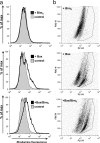
Bax expression causes mitochondrial hyperpolarization in yeast, and this effect is enhanced by BimS. Yeast strains containing BimS, Bax, or Bax/BimS were grown in the absence of tetracycline (to induce Bax expression) for 24 h. An untransformed yeast strain was used as control. Yeast cells were grown to an OD600 of 0.2 and incubated with 5 μM rhodamine123 for 30 min at 30°C. Cells were analyzed by flow cytometry. (a) Histograms showing rhodamine123 fluorescence. (b) Two-dimensional dot-plot of rhodamine123 versus forward scatter. Data are representative of six individual experiments.

Similar articles

Cited by

References

    1. Adams, J.M. 2003. Ways of dying: multiple pathways to apoptosis. Genes Dev. 17:2481–2495. - PubMed
    1. Cartron, P.F., H. Arokium, L. Oliver, K. Meflah, S. Manon, and F.M. Vallette. 2005. Distinct domains control the addressing and the insertion of Bax into mitochondria. J. Biol. Chem. 280:10587–10598. - PubMed
    1. Certo, M., G. Moore Vdel, M. Nishino, G. Wei, S. Korsmeyer, S.A. Armstrong, and A. Letai. 2006. Mitochondria primed by death signals determine cellular addiction to antiapoptotic BCL-2 family members. Cancer Cell. 9:351–365. - PubMed
    1. Chen, L., S.N. Willis, A. Wei, B.J. Smith, J.I. Fletcher, M.G. Hinds, P.M. Colman, C.L. Day, J.M. Adams, and D.C. Huang. 2005. Differential targeting of prosurvival Bcl-2 proteins by their BH3-only ligands allows complementary apoptotic function. Mol. Cell. 17:393–403. - PubMed
    1. Cory, S., and J.M. Adams. 2002. The Bcl2 family: regulators of the cellular life-or-death switch. Nat. Rev. Cancer. 2:647–656. - PubMed

Publication types

MeSH terms