Comparative Study
. 2007 Jun 11;204(6):1383-93.
doi: 10.1084/jem.20061205. Epub 2007 May 21.

Mice lacking NKCC1 are protected from development of bacteremia and hypothermic sepsis secondary to bacterial pneumonia

Affiliations
Comparative Study

Mice lacking NKCC1 are protected from development of bacteremia and hypothermic sepsis secondary to bacterial pneumonia

MyTrang Nguyen et al. J Exp Med. .

Abstract

The contribution of the Na(+)-K(+)-Cl(-) transporter (NKCC1) to fluid in ion transport and fluid secretion in the lung and in other secretory epithelia has been well established. Far less is known concerning the role of this cotransporter in the physiological response of the pulmonary system during acute inflammation. Here we show that mice lacking this transporter are protected against hypothermic sepsis and bacteremia developing as a result of Klebsiella pneumoniae infection in the lung. In contrast, this protection was not observed in NKCC1(-/-) mice with K. pneumoniae-induced peritonitis. Although overall recruitment of cells to the lungs was not altered, the number of cells present in the airways was increased in the NKCC1(-/-) animals. Despite this robust inflammatory response, the increase in vascular permeability observed in this acute inflammatory model was attenuated in the NKCC1(-/-) animals. Our studies suggest that NKCC1 plays a unique and untoward unrecognized role in acute inflammatory responses in the lung and that specific inhibition of this NKCC isoform could be beneficial in treatment of sepsis.


Figures
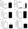
Figure 1.
Figure 1.
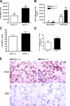
Inflammatory cells and cytokines present in BALF 48 h after infection with K. pneumoniae. (A) Total number of cells present in the airways and thus recovered by BAL was higher in the lungs of NKCC1−/− mice (n = 30) than NKCC1+/+ mice (n = 29). (B) Cells present in the BAL were stained and identified based on morphological criteria. There was a statistically significant difference in the number of neutrophils in NKCC1−/− mice compared with wild-type controls. Macrophages remained unchanged in both groups. TNF-α, IL-1β, IL-10, and IL-6 levels were determined in the lung homogenates by ELISA kits. NKCC1−/− mice exhibited increased levels of (C) TNF-α and (D) IL-1β, but the increased levels did not reach statistical significance compared with littermate controls. (E) IL-10 levels were significantly reduced in NKCC1−/− mice compared with NKCC1+/+ mice. (F) IL-6 levels were also reduced in NKCC1−/− mice in comparison to NKCC1+/+ mice, but the reduced levels were not significant. Data are expressed as mean ± SEM; n = 7–10 mice per group for cytokine assays. *, P < 0.05; **, P < 0.005. conc., concentration.
Figure 2.
Figure 2.
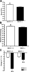
Colonization of the lung, bacteremia, and hypothermic sepsis after intertracheal infection of NKCC1−/− and control animals with K. pneumoniae. (A) Bacterial CFUs in the blood and (B) lung homogenates of NKCC1−/− mice (n = 7–8) were significantly lower than NKCC1+/+ mice (n = 10–14). (C) Core temperatures of NKCC1−/− and wild-type littermates were determined before K. pneumoniae infection and 24 and 48 h thereafter. No differences were observed between the groups before inoculation or 24 h postinfection. However, the temperature drop observed in NKCC1−/− mice was significantly less than that measured for the NKCC1+/+ mice 48 h postinfection. Values are shown as mean ± SEM; n = 38–43 mice per group. *, P < 0.05; **, P < 0.000005.
Figure 3.
Figure 3.
Oxidative burst and killing of K. pneumoniae by NKCC1+/+ and NKCC1−/− neutrophils. (A) After 0, 30, and 60 min of incubation with K. pneumoniae, neutrophils isolated from the bone marrow of mice lacking NKCC1 did not differ from wild-type neutrophils in their ability to kill K. pneumoniae ex vivo. (B) Similarly, no difference was observed in superoxide production elicited by stimulation of the NKCC1−/− and NKCC1+/+ neutrophils with PMA. Results are shown as mean ± SEM; n = 5 mice per group.
Figure 4.
Figure 4.
NKCC1 mRNA levels in lung and inflammatory cells. NKCC1 mRNA expression levels are significantly higher in the lung, an endothelial cell line (EOMA cell line, hemangioendothelioma origin, American Type Culture Collection no. CRL-2586), and primary lung endothelial cells (Primary Endo) relative to expression levels of zymosan-elicited neutrophils (Zym Neutrophils), bone marrow neutrophils (BM Neutrophils), BM mast cells (BMMC), and alveolar macrophages (Aveolar Mac). Data are expressed as mean ± SEM; n = 5 mice per group. *, , δ, P < 0.01 relative to alveolar macrophage mRNA. Statistical analysis was performed using one-way ANOVA with Tukey's Multiple Comparisons posttest.
Figure 5.
Figure 5.
Assessment of bacterial load, cellular infiltrates, and edema formation after induction of peritonitis with K. pneumoniae in NKCC1−/− and wild-type mice. (A) CFUs in the peritoneal lavage fluid of NKCC1+/+ and NKCC1−/− mice. Bacterial load was slightly lower in NKCC1−/− mice, but the reduced levels did not reach statistical significance (7–8 mice per group). (B) The total number of cells collected by peritoneal lavage was significantly decreased in NKCC1−/− mice compared with littermate controls (P = 0.04). (C) Differential cell counts were determined, based on morphological criteria, of cells present in the peritoneal lavage fluid of infected mice. A significant decrease was observed in the number of peritoneal macrophages in NKCC1-deficient mice compared with wild-type controls (P = 0.016). Emigrated neutrophils were present 1 h after K. pneumoniae infection. However, NKCC1-deficient mice showed no compromise in neutrophil emigration compared with NKCC+/+ mice. (D) Consistent with a less robust inflammatory response, edema formation was significantly reduced in NKCC1−/− mice compared with littermate controls. Values are shown as mean ± SEM; n = 15–18 mice per group. *, P < 0.05.
Figure 6.
Figure 6.
Examination of the response of NKCC1−/− and control animals in a sterile model of acute lung inflammation. The total number of cells in the BALF was enumerated (A), and populations of leukocytes were determined based on morphological criteria (B). The total number of cells in the BAL was significantly increased in LPS-challenged NKCC1−/− mice (P = 0.00007; n = 11) compared with the LPS-challenged NKCC1+/+ mice (n = 12). Differential cell counts showed that this increase was the result primarily of increased numbers of neutrophils (P = 0.0001) although a small but significant increase in the number of macrophages was also observed (P = 0.004) in NKCC1-deficient mice compared with wild-type controls. To determine whether this difference represented a change in sequestration of neutrophils in the lung or specifically the movement of the cells into the airway, lungs were harvested from similarly treated animals, and the level of MPO in the lung was determined (C). MPO levels in the lungs of NKCC1−/− mice (n = 11) were higher than in the wild-type controls (n = 12), but the increased levels did not reach statistical significance. To directly visualize differences in the location of inflammatory cells in the two mouse lines, lungs were fixed and sections were prepared from LPS and saline-treated animals 12 h after exposure. Images similar to those shown in E were scanned and analyzed using Image Scope viewing software, and cells present in the airways were enumerated in a blinded fashion. The number of neutrophils present in the airways of the NKCC1−/− lungs (n = 4) was higher than in the control animals (n = 4) (D). All values are expressed as mean ± SEM. *, P < 0.005 and **, P < 0.0005. Bars: (top) 50 μm; (bottom) 100 μm.
Figure 7.
Figure 7.
Inflammatory mediator levels in the lung homogenates of LPS-challenged NKCC1+/+ and NKCC1−/− mice. No significant difference was observed in the levels of TNF-α, KC, and MIP-2 between NKCC1+/+ and NKCC1−/− mice. However, NKCC1−/− mice (P = 0.05) exhibited slightly but significantly higher levels of IL-6 in comparison to littermate controls. Values are mean ± SEM from 11–12 mice per group. *, P < 0.05. conc., concentration.
Figure 8.
Figure 8.
Evaluation of the contribution of NKCC1 expression by hematopoietic and lung cells to LPS-elicited neutrophil recruitment. We first performed an experiment to determine whether a difference in neutrophil recruitment and localization would continue to be observed in mice exposed to lethal doses of irradiation. NKCC1−/− and NKCC1+/+ mice were lethally irradiated and reconstituted with genetically identical marrow (A). 6–8 wk after engraftment mice were challenged with LPS, the total cells in the BALF were enumerated, and the MPO content of the BAL and the lung was determined. As expected, we continued to observe an increase in the number of neutrophils present in the BAL, although no difference in the total number of cells recruited to the lung was apparent. To determine whether expression of NKCC1 by neutrophils contributes to the differential distribution of these cells, we generated a second cohort of mice. In this case, after irradiation, NKCC1−/− mice received marrow from wild-type animals (NKCC+/+→ NKCC−/−), whereas NKCC1+/+ mice received marrow from NKCC1−/− mice (NKCC−/−→ NKCC+/+) (B). Higher cell counts in the BALF and higher MPO levels in the cells collected by lavage corresponded with loss of NKCC1 expression in the recipient, not the donor marrow (NKCC+/+→ NKCC−/−, n = 4). Cell counts and MPO activity were significantly higher in these animals compared with wild-type animals in which the recipient expressed NKCC1 but was reconstituted with NKCC1−/− hematopoietic cells (NKCC−/−→ NKCC+/+, n = 6). Again, no difference was observed between the groups in MPO activity in the lung. All values are expressed as mean ± SEM. *, P < 0.05.
Figure 9.
Figure 9.
Western blot analysis of VCAM-1 after LPS stimulation. Lung lysates (100 μg of protein/lane) were subjected to Western blot analysis using polyclonal anti–mouse VCAM-1 antibody. GAPDH antibody was used as a loading control. Western blot analysis demonstrates an increase in VCAM-1 protein expression induced by LPS. However, the increased levels did not differ between NKCC1+/+ and NKCC1−/− mice.
Figure 10.
Figure 10.
Changes in the permeability of lung capillaries after exposure of mice to LPS. Evan's blue extravasation was significantly lower in NKCC1−/− mice (n = 7) than in NKCC1+/+ mice (n = 6) both 3 and 6 h after challenge. Similarly, a decrease in the wet-to-dry ratio was observed in the NKCC1−/− animals 3 h after LPS exposure (n = 25 NKCC1+/+, n = 18 NKCC1−/−). Data are expressed as mean ± SEM. *, #, P< 0.05.

Similar articles

Cited by

References

    1. Wheeler, A.P., and G.R. Bernard. 1999. Treating patients with severe sepsis. N. Engl. J. Med. 340:207–214. - PubMed
    1. Hudson, L.D., and K.P. Steinberg. 1999. Epidemiology of acute lung injury and ARDS. Chest. 116:74S–82S. - PubMed
    1. Hasleton, P.S., and T.E. Roberts. 1999. Adult respiratory distress syndrome—an update. Histopathology. 34:285–294. - PubMed
    1. Ali, J., W. Chernicki, and L.D. Wood. 1979. Effect of furosemide in canine low-pressure pulmonary edema. J. Clin. Invest. 64:1494–1504. - PMC - PubMed
    1. Prabhu, V.G., M. Keszler, and R. Dhanireddy. 1997. Pulmonary function changes after nebulised and intravenous frusemide in ventilated premature infants. Arch. Dis. Child. Fetal Neonatal Ed. 77:F32–F35. - PMC - PubMed

Publication types

MeSH terms

Substances