. 2007 Jun 11;204(6):1249-56.
doi: 10.1084/jem.20070301. Epub 2007 May 21.

Spontaneous hemolytic uremic syndrome triggered by complement factor H lacking surface recognition domains

Affiliations

Spontaneous hemolytic uremic syndrome triggered by complement factor H lacking surface recognition domains

Matthew C Pickering et al. J Exp Med. .

Abstract

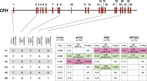
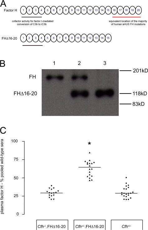
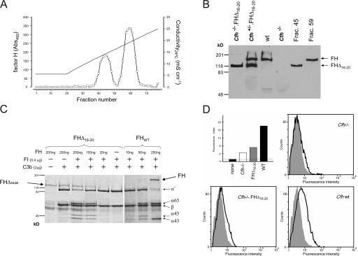
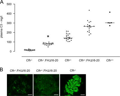
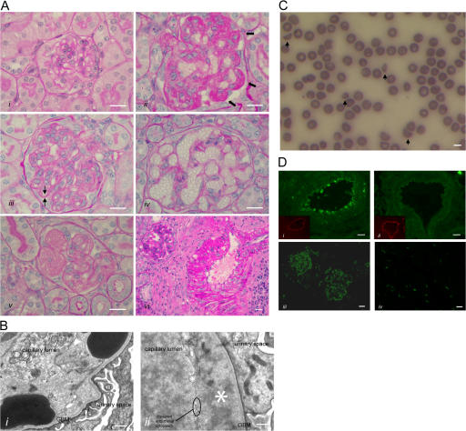
Factor H (FH) is an abundant serum glycoprotein that regulates the alternative pathway of complement-preventing uncontrolled plasma C3 activation and nonspecific damage to host tissues. Age-related macular degeneration (AMD), atypical hemolytic uremic syndrome (aHUS), and membranoproliferative glomerulonephritis type II (MPGN2) are associated with polymorphisms or mutations in the FH gene (Cfh), suggesting the existence of a genotype-phenotype relationship. Although AMD and MPGN2 share pathological similarities with the accumulation of complement-containing debris within the eye and kidney, respectively, aHUS is characterized by renal endothelial injury. This pathological distinction was reflected in our Cfh association analysis, which demonstrated that although AMD and MPGN2 share a Cfh at-risk haplotype, the haplotype for aHUS was unique. FH-deficient mice have uncontrolled plasma C3 activation and spontaneously develop MPGN2 but not aHUS. We show that these mice, transgenically expressing a mouse FH protein functionally equivalent to aHUS-associated human FH mutants, regulate C3 activation in plasma and spontaneously develop aHUS but not MPGN2. These animals represent the first model of aHUS and provide in vivo evidence that effective plasma C3 regulation and the defective control of complement activation on renal endothelium are the critical events in the molecular pathogenesis of FH-associated aHUS.


Figures

Figure 1.
Figure 1.
Association analysis of CFH haplotypes with aHUS, AMD, and MPGN2 within a single population. Schematic illustration of the CFH exon structure demonstrating the location of the five SNPs included in these studies. These SNPs represent a minimal informative set for genetic variation within the CFH gene. CFH haplotypes with a frequency >3% are shown. The frequency of each CFH haplotype was compared with the controls and the aHUS, AMD, and MPGN2 cohorts, and the p-values and the OR were calculated. Risk haplotypes are shaded red, whereas protective haplotypes are shaded in green.p-values were derived using the two-sided Fisher's exact test. OR and 95% confidence interval (95% CI) are shown. See Table S1 for individual CFH SNP allele frequencies in patients with these conditions. The nucleotide and amino acid numbering refer to the translation start site (A in ATG is +1; Met is +1), as recommended by the Human Genome Variation Society.
Figure 2.
Figure 2.
The development of Cfh−/−.FHΔ16-20 mice. (A) Schematic representation of the mouse FH protein and the mutant mouse FHΔ16-20 protein. SCR domains are numbered incrementally from the amino terminus. Complement regulatory domains are localized within SCR domains 1–4 (black line), whereas the equivalent location of the majority of aHUS-associated human mutations is within SCR domains 16–20 (red line). (B) Western blot of plasma probed with a polyclonal anti–mouse FH antibody from wild-type (lane 1), Cfh +/−.FHΔ16-20 (lane 2), and Cfh −/−.FHΔ16-20 mice (lane 3). The truncated mutant FHΔ16-20 protein runs at a lower molecular mass than the 150-kD full-length mouse protein. (C) Plasma FH levels in Cfh −/−.FHΔ16-20, Cfh +/−.FHΔ16-20, and Cfh +/− mice. Median FHΔ16-20 plasma levels quantified by ELISA in Cfh −/−.FHΔ16-20 mice were 29.3% pooled wild-type sera (range = 20.1–39.1%; n = 16), which were comparable to FH levels in Cfh +/− mice (median = 28.9%, range = 20.5–50.5%; n = 21; P > 0.05). In the Cfh +/−.FHΔ16-20 mice, total FH levels were 64.5% (range = 46.7–84.6%; n = 18), significantly higher than levels in either the Cfh −/−.FHΔ16-20 (P < 0.001) or the Cfh +/− (P < 0.001) mice. Horizontal bars denote median values. *, P < 0.001 for Cfh +/−.FHΔ16-20 mice versus all other groups using Bonferroni's multiple comparison test.
Figure 3.
Figure 3.
Functional characterization of FHΔ16-20. (A and B) Heparin binding assay. Cfh −/+.FHΔ16-20 mouse plasma was applied to a heparin–sepharose column, and the proteins bound to the column were eluted with a NaCl linear gradient (35–250 mM). Two protein peaks containing FH identified by ELISA (A) and Western blot analysis (B) showed that the mutant FHΔ16-20 protein eluted before FH, demonstrating that removal of SCR16-20 impairs binding of FH to heparin. The continuous line in A indicates conductivity. (C) Cofactor activity of FHΔ16-20 protein in the proteolysis of fluid-phase mouse C3b by factor I. Different concentrations of either purified FH or FHΔ16-20 protein were incubated with mouse C3b in the presence of factor I. Analysis of C3b proteolytic fragments on 8% SDS-PAGE gel under reducing conditions indicated that both proteins had factor I cofactor activity with the appearance of α chain fragments (α65 and α45/43). Protein fragments were visualized using Coomassie blue staining. (D) HUVEC binding assays (background level indicated by the horizontal line; top left). HUVECs were incubated with 100 μl EDTA plasma dialyzed against 0.5× PBS (137 mM NaCl, 10 mM phosphate, 2.7 mM KCl, pH 7.4). Bound FH or FHΔ16-20 were detected using a rabbit anti–mouse FH antibody and a goat anti–rabbit Alexa Fluor 488–conjugated antibody. Alexa Fluor 488–conjugated isotype-matched antibody was used as a control (shaded area). The fluorescence index was calculated by multiplying the mean fluorescent intensity by the percentage of cells staining positive for FH (bold line). These analyses demonstrated that the mutant FHΔ16-20 protein has a markedly impaired ability to bind to HUVECs in comparison with wild-type protein.
Figure 4.
Figure 4.
Plasma and glomerular C3 regulation in Cfh−/−.FHΔ16-20 mice. (A) Plasma C3 levels in Cfh −/−.FHΔ16-20 mice. Median plasma C3 levels in the Cfh −/−.FHΔ16-20 mice were 79.5 mg/liter (range = 46.1–95.9 mg/liter; n = 16), which was significantly higher than the levels seen in the Cfh −/− mice (median = 14.3 mg/liter, range = 7.9–23.2 mg/liter; n = 9; P < 0.05). C3 levels were also significantly higher in the Cfh +/−.FHΔ16-20 mice compared with Cfh +/− mice (medians = 264.6 vs. 142 mg/liter, respectively; P < 0.001) and did not differ from the levels seen in wild-type animals. Horizontal bars denote median values. *, P < 0.05 for Cfh −/−.FHΔ16-20 mice versus all other groups using Bonferroni's multiple comparison test. (B) Glomerular C3 staining in 3-wk-old Cfh −/−.FHΔ16-20, Cfh +/−.FHΔ16-20, and Cfh −/− mice. The striking GBM linear C3 staining pattern seen in the glomeruli of the Cfh −/− mice was not evident in either Cfh −/−.FHΔ16-20 or Cfh +/−.FHΔ16-20 mice, consistent with the ability of FHΔ16-20 to prevent GBM C3 deposition. Although no staining was detected in the glomeruli of Cfh +/−.FHΔ16-20 animals, a granular mesangial C3 staining pattern was evident in Cfh −/−.FHΔ16-20 mice. Bar, 10 μm.
Figure 5.
Figure 5.
HUS in Cfh−/−.FHΔ16-20 mice. (A) Renal histology in Cfh −/−.FHΔ16-20 mice. Normal glomerulus from a 2-mo-old Cfh +/−.FHΔ16-20 mouse (i), and light microscopic features of thrombotic microangiopathy in Cfh −/−.FHΔ16-20 mice (ii–vi). These included glomerular microthrombi (ii, arrows), capillary wall double contours (iii, arrows), formation of capillary microaneurysms (iv), and mesangiolysis (v). Inflammatory changes were also seen within glomerular arteries (vi). Bar, 10 μm. (B) Electron microscopy revealed characteristic ultrastructural changes of thrombotic microangiopathy. Erythrocytes beneath disrupted endothelium and in direct contact with the GBM (i) and endothelial disruption with subendothelial accumulation of flocculent material (ii, asterisk). Note the absence of GBM electron-dense deposits, an ultrastructural feature of MPGN2 that is normally evident at this age in Cfh −/− mice (reference 12). Bar, 500 nm. (C) Peripheral blood smear from a Cfh −/−.FHΔ16-20 mouse with hematuria. Evidence of red-cell fragmentation is seen (arrows). Bar, 5 μm. (D) Renal C3 staining. C3 deposition along the endothelium and within the smooth muscle of renal arteries was present in the Cfh −/−.FHΔ16-20 (i) but not the Cfh +/−.FHΔ16-20 (ii) mice. Insets represent the staining of mouse endothelium with anti-CD31 (platelet/endothelial cell adhesion molecule). Mesangial and capillary wall C3 staining in a Cfh −/−.FHΔ16-20 mouse with HUS (iii) in contrast to an absence of abnormal glomerular C3 staining in an age-matched Cfh +/−.FHΔ16-20 mouse (iv). No abnormal renal IgG staining was present in either the Cfh −/−.FHΔ16-20 or the Cfh +/−.FHΔ16-20 mice (not depicted). Bar, 10 μm.

References

    1. Scholl, H.P., M. Fleckenstein, P.C. Issa, C. Keilhauer, F.G. Holz, and B.H. Weber. 2007. An update on the genetics of age-related macular degeneration. Mol. Vis. 13:196–205. - PMC - PubMed
    1. Caprioli, J., P. Bettinaglio, P.F. Zipfel, B. Amadei, E. Daina, S. Gamba, C. Skerka, N. Marziliano, G. Remuzzi, and M. Noris. 2001. The molecular basis of familial hemolytic uremic syndrome: mutation analysis of factor H gene reveals a hot spot in short consensus repeat 20. J. Am. Soc. Nephrol. 12:297–307. - PubMed
    1. Perez-Caballero, D., C. Gonzalez-Rubio, M.E. Gallardo, M. Vera, M. Lopez-Trascasa, S. Rodriguez de Cordoba, and P. Sanchez-Corral. 2001. Clustering of missense mutations in the C-terminal region of factor H in atypical hemolytic uremic syndrome. Am. J. Hum. Genet. 68:478–484. - PMC - PubMed
    1. Richards, A., M.R. Buddles, R.L. Donne, B.S. Kaplan, E. Kirk, M.C. Venning, C.L. Tielemans, J.A. Goodship, and T.H. Goodship. 2001. Factor H mutations in hemolytic uremic syndrome cluster in exons 18-20, a domain important for host cell recognition. Am. J. Hum. Genet. 68:485–490. - PMC - PubMed
    1. Warwicker, P., T.H. Goodship, R.L. Donne, Y. Pirson, A. Nicholls, R.M. Ward, P. Turnpenny, and J.A. Goodship. 1998. Genetic studies into inherited and sporadic hemolytic uremic syndrome. Kidney Int. 53:836–844. - PubMed

Publication types