. 2007 Mar;13(3):435-45.
doi: 10.1089/ten.2006.0182.

Structural bone allograft combined with genetically engineered mesenchymal stem cells as a novel platform for bone tissue engineering

Affiliations

Structural bone allograft combined with genetically engineered mesenchymal stem cells as a novel platform for bone tissue engineering

Chao Xie et al. Tissue Eng. 2007 Mar.

Abstract

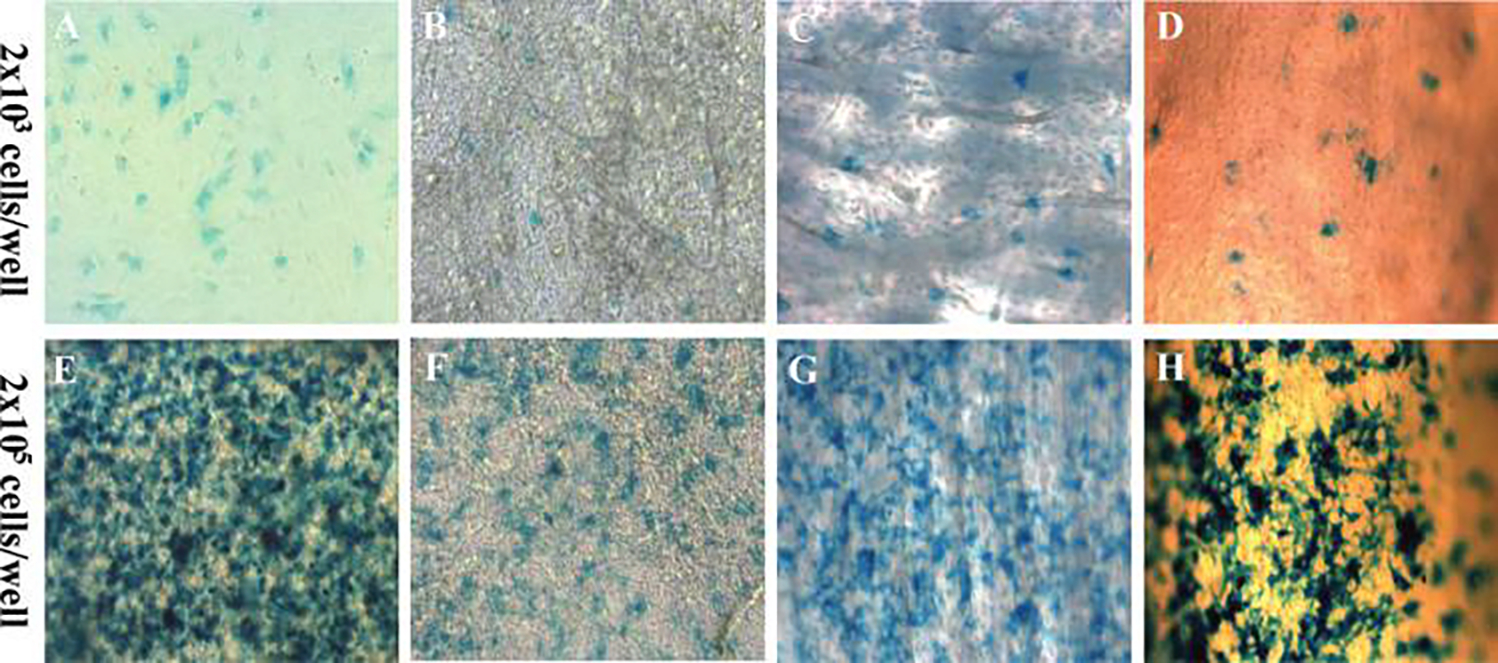
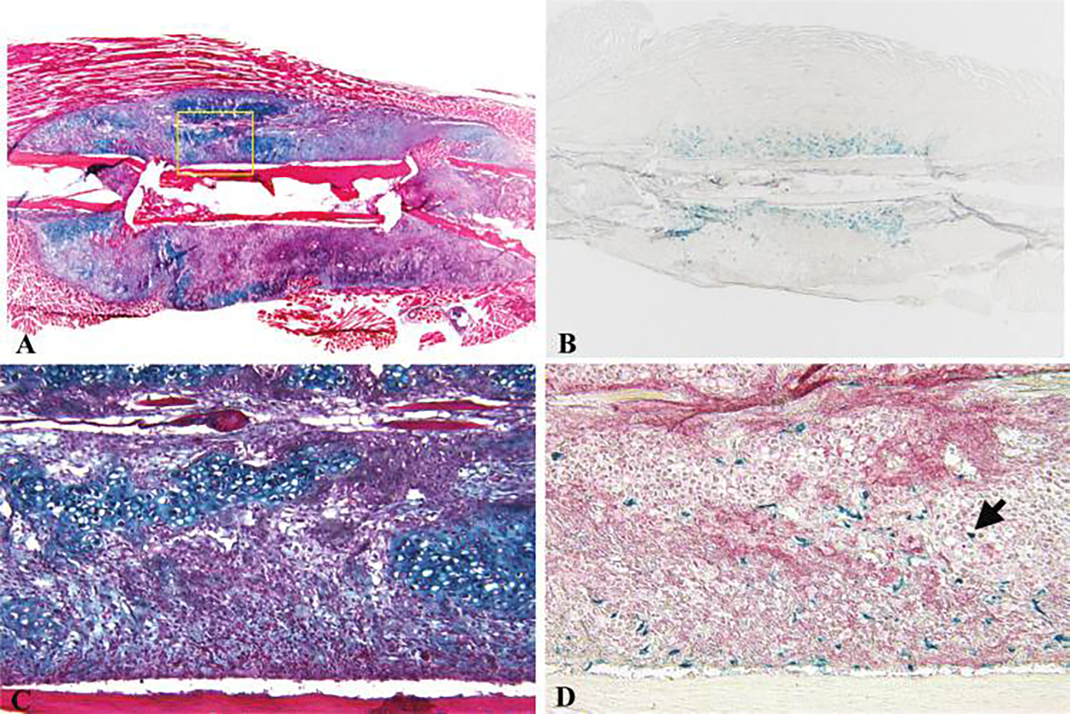
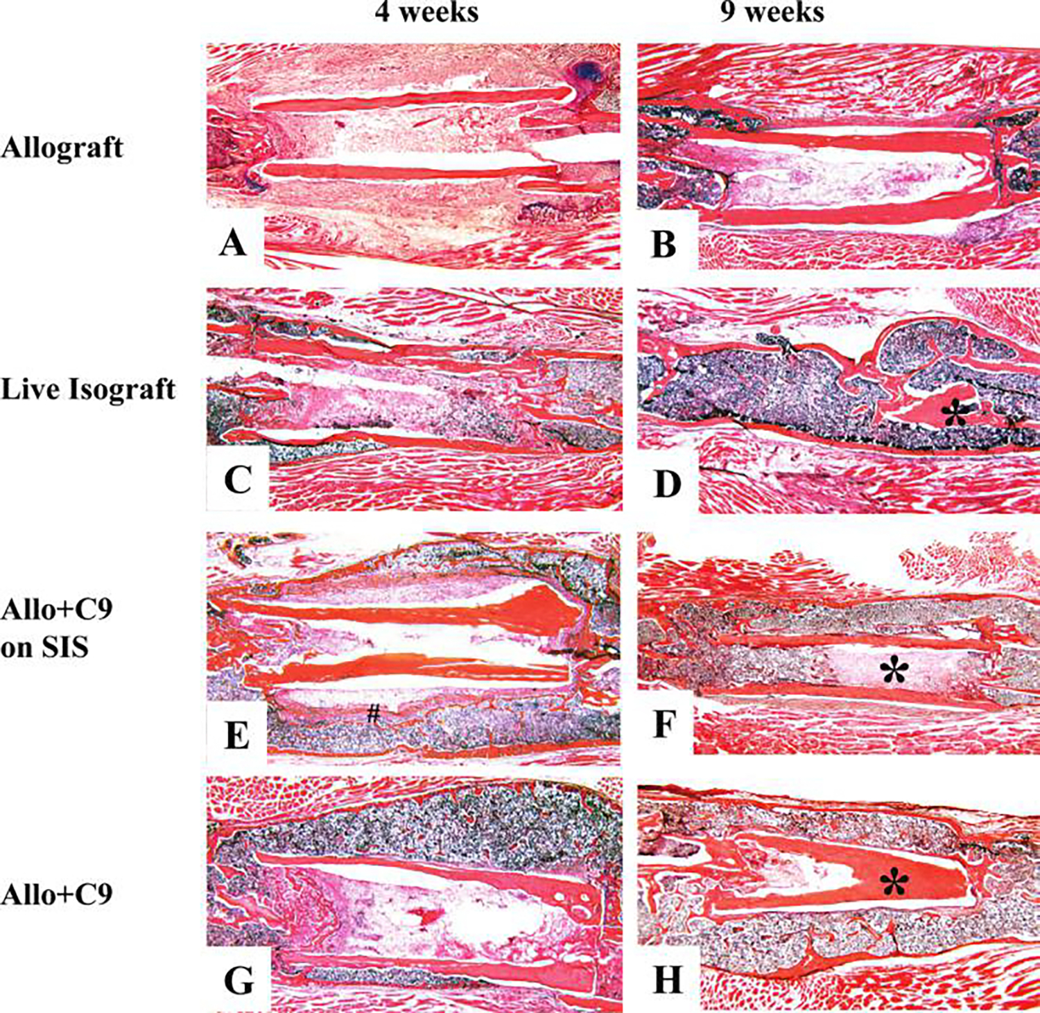
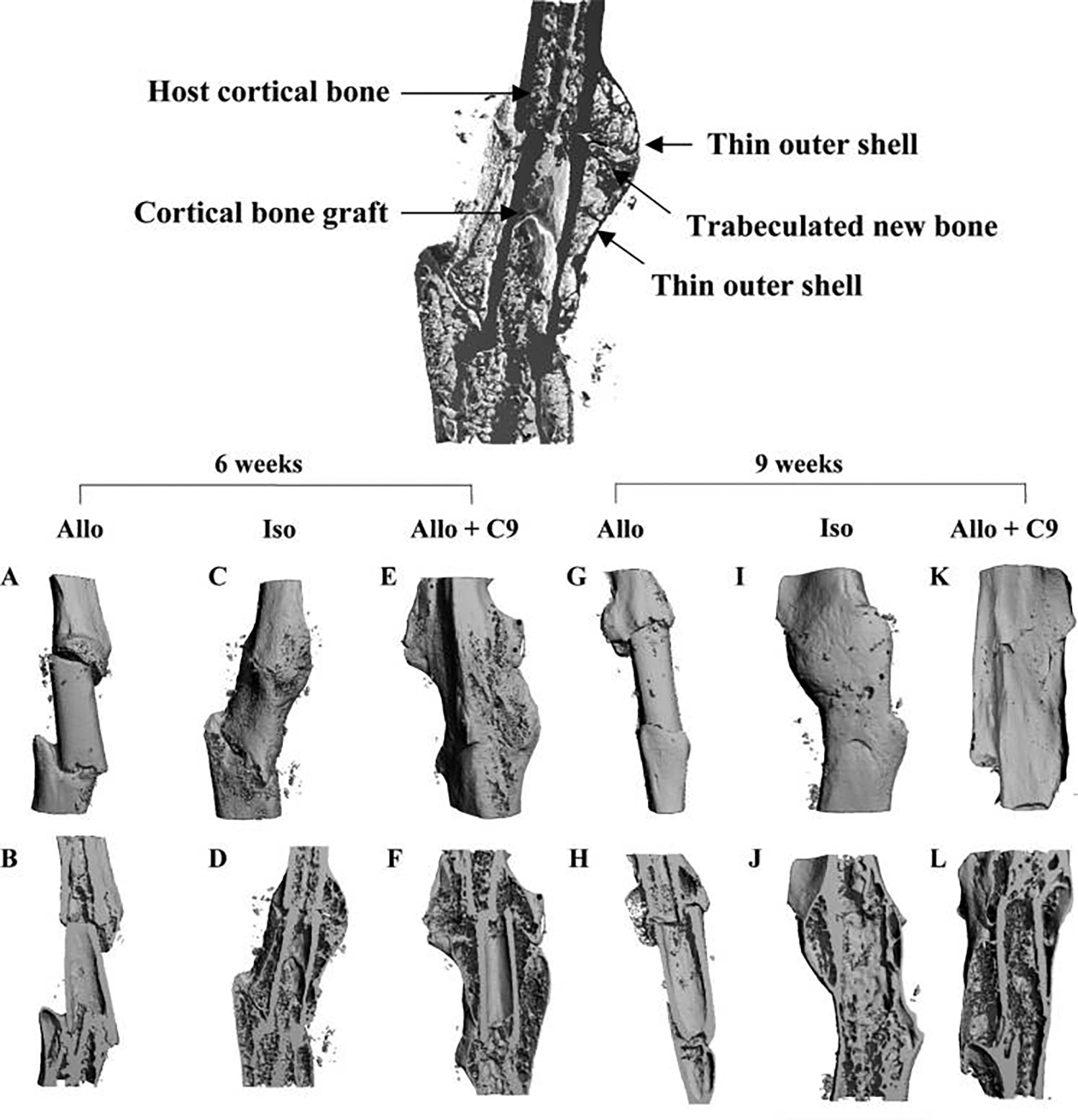
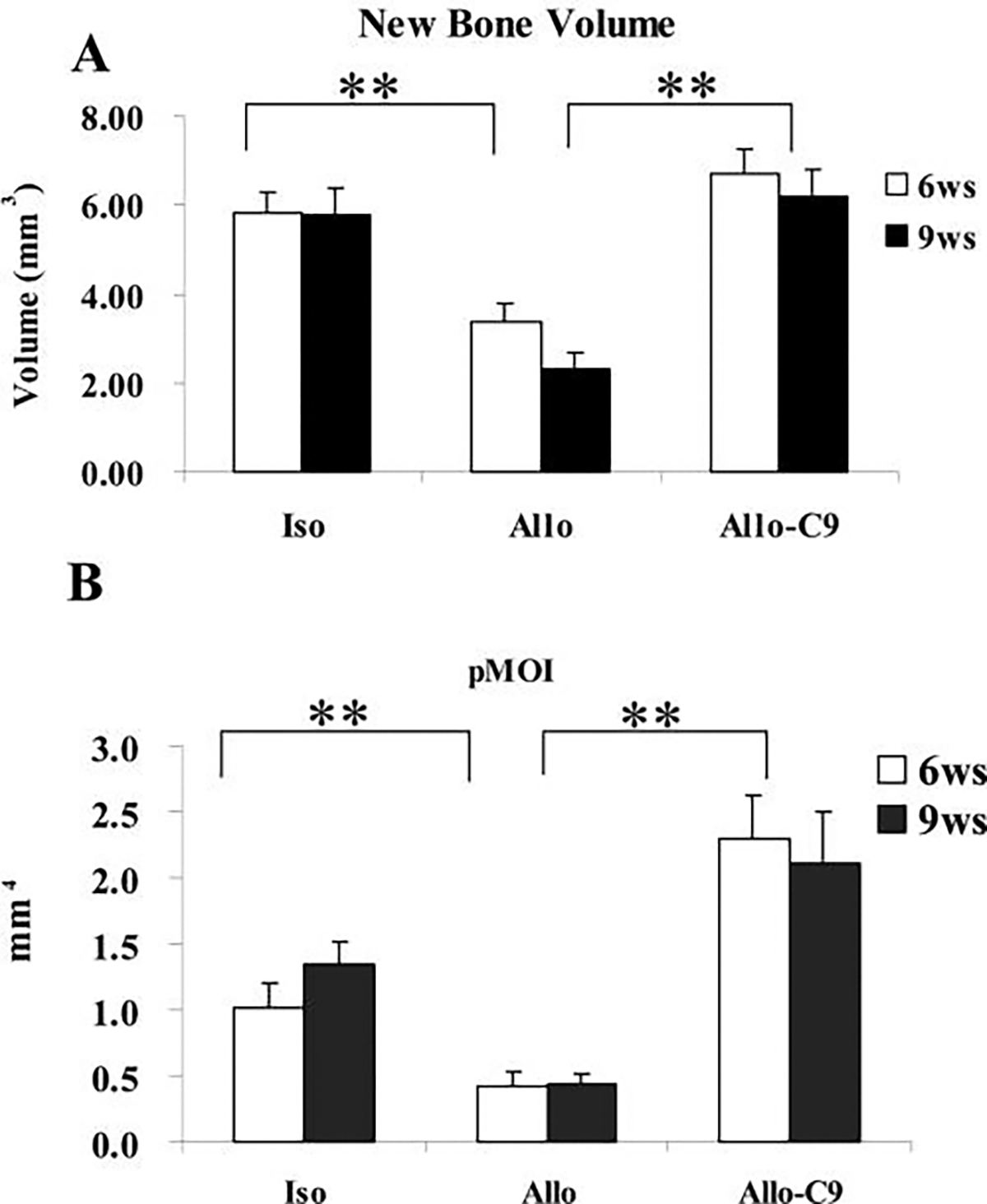
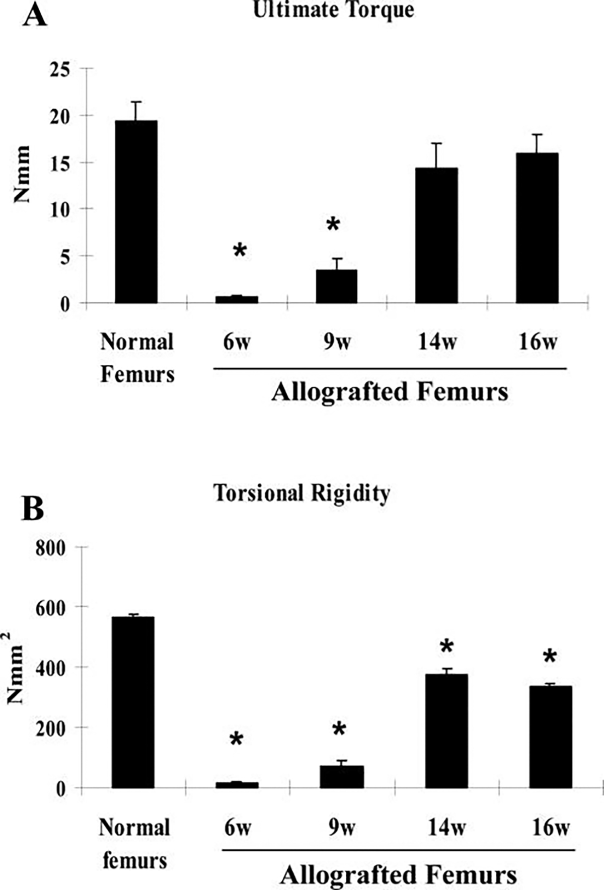
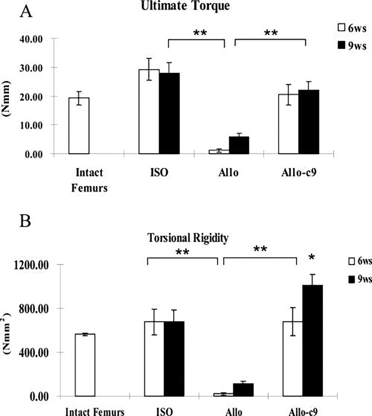
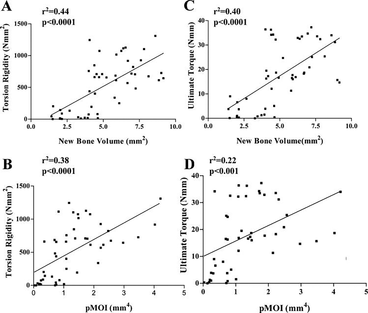
The presence of live periosteal progenitor cells on the surface of bone autografts confers better healing than devitalized allograft. We have previously demonstrated in a murine 4 mm segmental femoral bone-grafting model that live periosteum produces robust endochondral and intramembraneous bone formation that is essential for effective healing and neovascularization of structural bone grafts. To the end of engineering a live pseudo-periosteum that could induce a similar response onto devitalized bone allograft, we seeded a mesenchymal stem cell line stably transfected with human bone morphogenic protein-2/beta-galactosidase (C9) onto devitalized bone allografts or onto a membranous small intestinal submucosa scaffold that was wrapped around the allograft. Histology showed that C9-coated allografts displayed early cartilaginous tissue formation at day 7. By 6 and 9 weeks, a new cortical shell was found bridging the segmental defect that united the host bones. Biomechanical testing showed that C9-coated allografts displayed torsional strength and stiffness equivalent to intact femurs at 6 weeks and superior to live isografts at 9 weeks. Volumetric and histomorphometric micro-computed tomography analyses demonstrated a 2-fold increase in new bone formation around C9-coated allografts, which resulted in a substantial increase in polar moment of inertia (pMOI) due to the formation of new cortical shell around the allografts. Positive correlations between biomechanics and new bone volume and pMOI were found, suggesting that the biomechanical function of the grafted femur relates to both morphological parameters. C9-coated allograft also exhibited slower resorption of the graft cortex at 9 weeks than live isograft. Both new bone formation and the persistent allograft likely contributed to the improved biomechanics of C9-coated allograft. Taken together, we propose a novel strategy to combine structural bone allograft with genetically engineered mesenchymal stem cells as a novel platform for bone tissue engineering.


Figures

FIG. 1.
FIG. 1.
Culture of C9 mesenchymal stem cells on scaffold or bone. Bone morphogenic protein-2-producing mesenchymal stem cells (C9) were seeded on plastic surface (A&E), small intestinal submucosa membrane (B&F), bovine bone wafer (C&G), or murine bone allograft (D&H) at the indicated seeding density. Cells were stained using X-galactosidase to determine the resulting seeding density. Color images available online at www.liebertpub.com/ten.
FIG. 2.
FIG. 2.
Histologic healing of C9-coated allografts. C9 cells were directly seeded on an allograft and used to repair a 4-mm segmental bone defect in a mouse femur. Alcian blue and hematoxylin and eosin staining sections demonstrated direct induction of early cartilaginous callus on devitalized bone allograft in C9-coated allografts 7 days post-surgery (A&C). X-galactosidase staining in an adjacent section showed numerous β-galactosidase-positive C9 cells within the early periosteal callus (B&D). Alkaline phosphatase staining of the same section showed differentiated alkaline phosphatase–positive mesenchymal cells around the bone allograft (D). (Arrow indicates β-galactosidase-positive chondrocytes.) Color images available online at www.liebertpub.com/ten.
FIG. 3.
FIG. 3.
Induction of periosteal-like new bone callus in C9-coated allografts (Allo + C9). C9 cells were directly seeded on bone allograft or on small intestinal submucosa (SIS) membrane wrapped around the allograft. The cellularized allograft was used to repair a 4 mm segmental defect in the mouse femur. Representative histological sections from allograft (A&B), live isograft (C&D), allograft wrapped with C9 cells pre-seeded on SIS membrane (E&F), or allograft with direct C9 cell seeding (G&H) were obtained at 4 and 9 weeks post-grafting. The absence of periosteal bone formation associated with poor graft incorporation is evident in allograft 4 and 9 weeks post-surgery. In contrast, Allo + C9 produced a contiguous new bone callus with an outer cortical shell at 4 and 9 weeks post-surgery, similar to live isograft at 4 weeks. (* indicates transplanted bone graft, # indicates the residues of SIS). Color images available online at www.liebertpub.com/ten.
FIG. 4.
FIG. 4.
Three-dimensional micro-computed tomography ()imaging of C9-coated allografts (Allo + C9). Image at top center illustrates the structure layers of a healing isografted femur (Iso). Representative micro-CT images were obtained from grafted femurs 6 and 9 weeks post-surgery. Limited bone formation was observed in allograft (Allo) (A&B, G&H). In contrast, extensive new bone formation across bone graft was shown in intact live bone Iso at 6 weeks (C&D). By 9 weeks, live bone graft cortex was resorbed, leaving the outer shell as the major load-bearing structure for the healing bone (I&J). In Allo + C9, periosteal bone formation was restored, which resulted in a contiguous new cortical shell at 6 and 9 weeks post-surgery (E&F, K&L). In addition, graft resorption was markedly delayed to allow the persistent allograft and the outer cortical shell to form a contiguous load-bearing structure.
FIG. 5.
FIG. 5.
C9-coated allografts (Allo-C9) demonstrate a significant increase in bone formation. New bone volume around allografts (Allo) (A) and average cross-sectional polar moment of inertia of the grafted region (B) were markedly greater in Allo-C9. Data shown are means ± standard errors of the mean. * p < 0.05 (n ≥ 8). Iso, isograft.
FIG. 6.
FIG. 6.
Torsional testing indicates inferior biomechanics of allografted femurs. Torsional testing was performed to examine the mechanical properties of allografted femurs at 6, 9, 14, and 16 weeks. Normal intact femurs were used as controls. Each group included 6samples. Data shown are means ± standard errors of the mean. * p < 0.05.
FIG. 7.
FIG. 7.
C9-coated allografts demonstrate a significant increase in biomechanical strength. Torsional testing was performed to determine the mechanical properties of allografts (Allo), live isografts (ISO), and C9-cell-treated allografts (Allo-C9). Ultimate torque (A) and torsional rigidity (B) were markedly greater in Allo-C9 than in devitalized Allo alone and were similar to those of the ISO. Normal intact femurs were used as controls. Data shown are means ± standard errors of the mean. * p < 0.05 (n ≥ 8).
FIG. 8.
FIG. 8.
Significant correlation between femur biomechanics and new bone volume and polar moment of inertia. Linear regression analyses were performed to determine the relationship between torsional rigidity, ultimate torque, and new bone volume, or pMOI, of allografts, isografts, and C9-coated allografts. The coefficients of determination (r2) for the correlation between torsional rigidity and new bone volume or pMOI were 0.44 and 0.38, respectively (n = 48, p < 0.001). The r2 for the correlation between ultimate torque and new bone volume or pMOI were 0.40 and 0.22, respectively (n = 48, p < 0.001).

Similar articles

Cited by

References

    1. Burchardt H 1983. The biology of bone graft repair. Clin Orthop 28–42. - PubMed
    1. Burchardt H 1987. Biology of bone transplantation. Orthop Clin North Am 18:187–196. - PubMed
    1. Lord CF, Gebhardt MC, Tomford WW, and Mankin HJ 1988. Infection in bone allografts. Incidence, nature, and treatment. J Bone Joint Surg Am 70:369–376. - PubMed
    1. Berrey BH Jr., Lord CF, Gebhardt MC, and Mankin HJ 1990. Fractures of allografts. Frequency, treatment, and end-results. J Bone Joint Surg Am 72:825–833. - PubMed
    1. Enneking WF, and Mindell ER 1991. Observations on massive retrieved human allografts. J Bone Joint Surg Am 73:1123–1142. - PubMed

Publication types