. 2007 May 29;104(22):9319-24.
doi: 10.1073/pnas.0701212104. Epub 2007 May 22.

Canonical Wnt signaling functions in second heart field to promote right ventricular growth

Affiliations

Canonical Wnt signaling functions in second heart field to promote right ventricular growth

Di Ai et al. Proc Natl Acad Sci U S A. .

Abstract

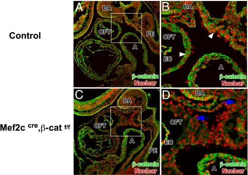
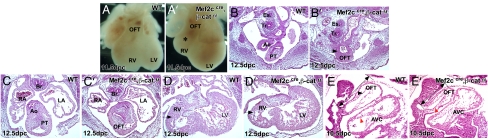
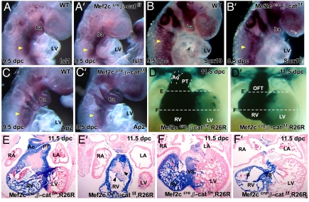
The second heart field (SHF), progenitor cells that are initially sequestered outside the heart, migrates into the heart and gives rise to endocardium, myocardium, and smooth muscle. Because of its distinct developmental history, the SHF is likely subjected to different signals from that of the first heart field. Previous experiments revealed that canonical Wnt signaling negatively regulated first heart field specification. We inactivated the obligate canonical Wnt effector beta-catenin using a beta-catenin conditional null allele and the Mef2c AHF cre driver that directs cre activity specifically in SHF. We also expressed a stabilized form of beta-catenin to model continuous Wnt signaling in SHF. Our data indicate that Wnt signaling acts in a positive fashion to promote right ventricular and interventricular myocardial expansion. Cyclin D2 and Tgfbeta2 expression was drastically reduced in beta-catenin loss-of-function mutants, indicating that Wnt signaling is required for patterning and expansion of SHF derivatives. Our findings reveal that Wnt signaling plays a major positive role in promoting growth and diversification of SHF precursors into right ventricular and interventricular myocardium.


Conflict of interest statement

The authors declare no conflict of interest.

Figures

Fig. 1.
Fig. 1.
Mef2ccre induced β-catenin deletion in SHF. Immunofluorescent staining of β-catenin in 9.5 dpc sagittal sections of control (A and B) and Mef2ccre; β-cateninflox mutant (C and D) embryos revealing membrane-localized β-catenin staining in ectoderm and endoderm as well as nuclear β-catenin in OFT myocardium, posterior pericardial mesothelium, and pharyngeal mesenchyme (denoted by white arrowheads in B). In the Mef2ccre; β-cateninflox mutant there is reduced β-catenin expression in pharyngeal mesenchyme and OFT myocardium, but not endocardium, due to mosaic activity of Mef2ccre in OFT endocardium (decreased β-catenin denoted by blue arrows). A, atrium; BA, branchial arch; EC, endocardium; PE, pharyngeal endoderm.
Fig. 2.
Fig. 2.
Phenotype of Mef2ccre; β-catenin mutant embryos. Shown are morphology and histology of 10.5–12.5 dpc β-catenin loss-of-function mutant hearts showing severe RV hypoplasia (∗) (A′) compared with control littermate (A). Transverse section of 12.5 dpc (B, B′, C, C′, D, and D′) and sagittal section of 10.5 dpc (E and E′) control and β-catenin loss-of-function mutant hearts reveal severe cardiac abnormalities: OFT septation defect in the distal OFT (B and B′, arrowheads) but not in more proximal OFT (C and C′), and hypoplasia of RV myocardium (D and D′, arrow) and short OFT (E and E′, double-headed arrows). Endocardium is denoted by arrowheads. PT, pulmonary trunk; LV, left ventricle; RV, right ventricle; RA, right atrium; LA, left atrium; Es, esophagus, Tr, Trachea, Br, bronchi.
Fig. 3.
Fig. 3.
Marker analysis of SHF and CNC and Mef2ccre lineage tracing in Mef2ccre; β-cateninflox mutants. In situ hybridizations of Isl1 (A and A′), Sox10 (B and B′), and Ap2 (C and C′) at 9.5 dpc indicated that SHF and CNC were correctly specified in Mef2ccre; β-catenin conditional mutant embryos (yellow arrowheads denote hybridization signal). Mef2ccre lineage tracing (D and D′) and transverse section (E, E′, F, and F′) in wild type and β-catenin mutant revealed a severe deficiency in the Mef2c lineage in Mef2ccre; β-catenin conditional mutant embryos. LV, left ventricle; RV, right ventricle; RA, right atrium; LA, left atrium; AO, aorta; PT, pulmonary trunk; Ba, branchial arch.
Fig. 4.
Fig. 4.
Marker analysis of OFT myocardium in Mef2ccre; β-cateninflox mutants. In situ hybridization with Cyclin D2 (A–D), Tgfβ2 (E and F), and Smarcd3 (G and H) in control and Mef2ccre; β-cateninflox mutants at 9.5 dpc. Pharyngeal mesenchyme is denoted by yellow arrowheads, and OFT is denoted by black arrowheads. (I and J) Sagittal sections of phospho-histone H3 immunostaining at 9.5 dpc. Arrowheads denote PH3 signal. lv, left ventricle; ba, brachial arch.
Fig. 5.
Fig. 5.
Phenotype of Mef2ccre; β-cateninEx3flox mutant embryos. Morphology and serial, transverse sections of 9.5 dpc wild-type (A, C, and E) and Mef2ccre; β-cateninEx3flox mutant embryos (B, D, and F) showing short but more dense and hypercellular OFT myocardium (denoted by black arrows). nt, neural tube; ba, branchial arch; LV, left ventricle; ph, pharynx.
Fig. 6.
Fig. 6.
Marker analysis in β-catenin loss-of-function and gain-of-function mutant embryos. (A–L) Analysis of markers for SHF and differentiated myocardium in 9.5-dpc embryos. Genotypes and markers are labeled. Pharyngeal mesenchyme is denoted by yellow arrowheads, and OFT is denoted by black arrowheads. OFT is outlined by a dotted line in G–I for clarity. (M) Model depicting the proposed role of canonical Wnt signaling in SHF. Based on loss- and gain-of-function experiments, canonical Wnt signaling promotes Bmp4 expression while concurrently acting to restrict Fgf10 in the SHF. The dotted line for Fgf10 repression indicates that this may be a minor function for Wnt signaling in the SHF because the data supporting this inhibitory function were observed only in the gain-of-function experiment. In the SHF-derived myocardium of the OFT, RV, and interventricular septum (IVS), canonical Wnt signaling regulates Bmp4 and promotes cell proliferation through regulation of Cyclin D. ba, branchial arch; RV, right ventricle; lv and LV, left ventricle; oc, otic capsule.

Similar articles

Cited by

References

    1. Meilhac SM, Esner M, Kerszberg M, Moss JE, Buckingham ME. J Cell Biol. 2004;164:97–109. - PMC - PubMed
    1. Meilhac SM, Esner M, Kelly RG, Nicolas JF, Buckingham ME. Dev Cell. 2004;6:685–698. - PubMed
    1. Waldo KL, Hutson MR, Ward CC, Zdanowicz M, Stadt HA, Kumiski D, Abu-Issa R, Kirby ML. Dev Biol. 2005;281:78–90. - PubMed
    1. Cai CL, Liang X, Shi Y, Chu PH, Pfaff SL, Chen J, Evans S. Dev Cell. 2003;5:877–889. - PMC - PubMed
    1. Buckingham M, Meilhac S, Zaffran S. Nat Rev Genet. 2005;6:826–835. - PubMed

Publication types

MeSH terms