. 2007 Aug;15(8):1512-21.
doi: 10.1038/sj.mt.6300207. Epub 2007 May 22.

Proteolytic disassembly is a critical determinant for reovirus oncolysis

Affiliations

Proteolytic disassembly is a critical determinant for reovirus oncolysis

Tommy Alain et al. Mol Ther. 2007 Aug.

Abstract

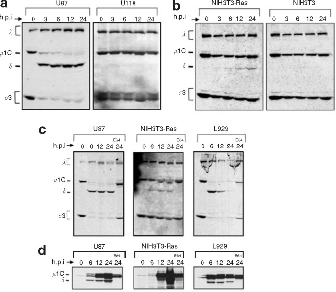
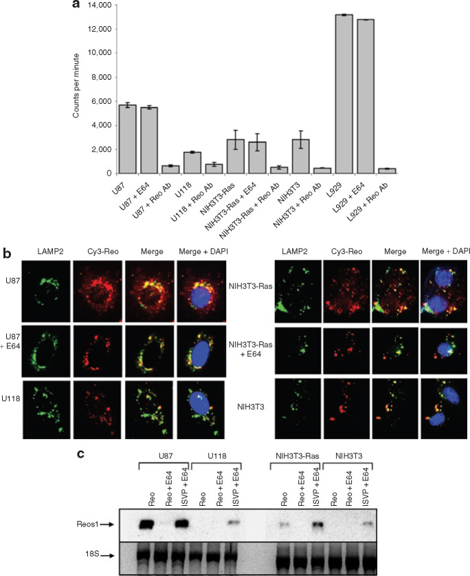
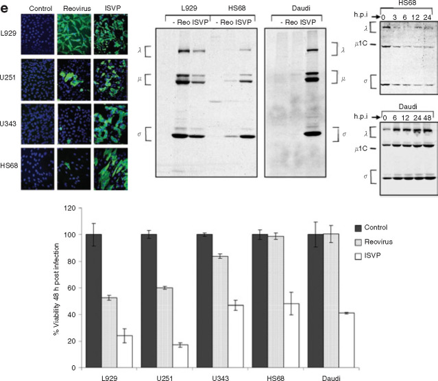
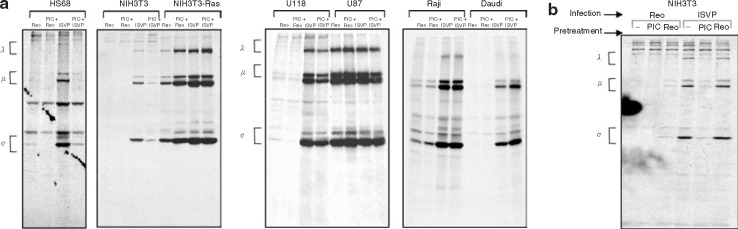
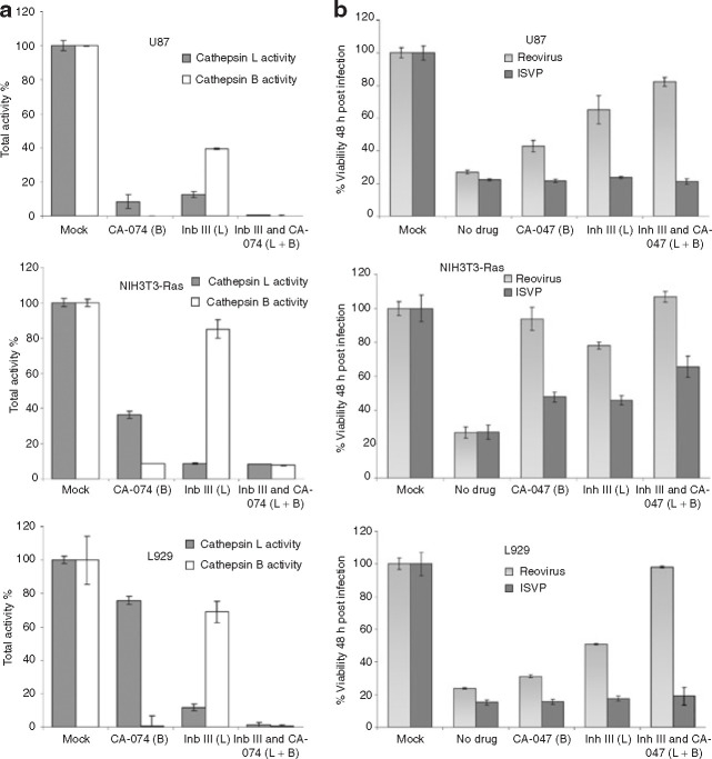
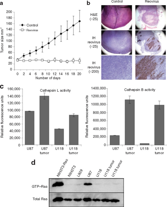
Mammalian ortheoreoviruses are currently being investigated as novel cancer therapeutics, but the cellular mechanisms that regulate susceptibility to reovirus oncolysis remain poorly understood. In this study, we present evidence that virion disassembly is a key determinant of reovirus oncolysis. To penetrate cell membranes and initiate infection, the outermost capsid proteins of reovirus must be proteolyzed to generate a disassembled particle called an infectious subviral particle (ISVP). In fibroblasts, this process is mediated by the endo/lysosomal proteases cathepsins B and L. We have analyzed the early events of infection in reovirus-susceptible and -resistant cells. We find that, in contrast to susceptible glioma cells and Ras-transformed NIH3T3 cells, reovirus-resistant cancer cells and untransformed NIH3T3 cells restrict virion uncoating and subsequent gene expression. Disassembly-restrictive cells support reovirus infection, as in vitro-generated ISVPs establish productive infection, and pretreatment with poly(I:C) does not prevent infection in cancer cells. We find that the level of active cathepsin B and L is increased in tumors and that disassembly-restrictive glioma cells support reovirus oncolysis when grown as a tumor in vivo. Together, these results provide a model in which proteolytic disassembly of reovirus is a critical determinant of susceptibility to reovirus oncolysis.


Figures

Figure 1
Figure 1
Evaluation of the proteolytic processing of reovirus in susceptible and resistant cell lines. (a) [35S]-labeled reovirus particles at 25,000 counts per minute were exposed to the glioma cell lines U87 and U118 as well as (b) Ras-transformed NIH3T3 (NIH3T3-Ras), and empty vector pBabe NIH3T3 (NIH3T3). Radiolabeled virus was allowed to bind at 4°C for 1 hour and then cells were returned to 37°C; cells were subsequently lysed at the defined time point after infection. Lysates were cleared of debris by centrifugation, and supernatants were submitted to sodium dodecyl sulfate polyacrylamide gel electrophoresis SDS-PAGE followed by autoradiography. (c) Reovirus proteolysis in susceptible cells treated with the E64 protease inhibitor (Sigma) in vitro and processed as described. Cells were exposed to 100 μmol/l of E64 for 1 hour before reovirus infection. After binding, E64 was again added to the cells to a final concentration of 100 μmol/l. (d) Western blot using a specific anti-μ1 reovirus antibody that detects both μ1C and δ was performed on a replicate of the experiment performed in c but using non-radioactive reovirions. h.p.i., hours post infection.
Figure 2
Figure 2
Assessment of reovirus binding, internalization, and viral transcription in susceptible and resistant cell lines. (a) [35S]-labeled reovirus was allowed to bind on cells in minimal medium for 1 hour at 4°C. Cells were washed twice in phosphate-buffered saline and lysed in a sonification buffer. Samples were subjected to scintillation counting, which was performed in triplicate. (b) Cy3-labeled reovirus at a multiplicity (MOI) of infection of 5,000 was added for 24 hours to cells grown on glass slides, after which cells were fixed in 4% formaldehyde. Immunofluorescence with the lysosomal marker lysosomal associated membrane protein 2 (LAMP2) antibody followed by secondary fluorescein isothiocyanate (FITC) was performed; slides were then mounted with a 4′,6-diamidino-2-phenylindole (DAPI) mounting medium (VECTOR) and photographed by multiple acquisition with a fluorescent Zeiss microscope (magnification ×400). (c) Total RNA from cells were purified using the RNeasy RNA extraction kit; equal RNA amounts were subjected to Northern blotting using a digoxygenin-labeled probe against the positive strand of reovirus S1 transcripts. ISVP, infectious subviral particle.
Figure 3
Figure 3
Infection of susceptible and resistant cell lines by proteolytically processed reovirus particles (infectious subviral particle or ISVPs). (a) Cells with or without treatment with the protease inhibitor E64 (100 μmol/l) 1 hour before reovirus exposure were pulse-labeled with [35S]methionine for 6 hours at 18 hours post infection (h.p.i.) with an MOI of 20 of reovirus or ISVPs (reovirions processed in 200 μg/ml of chymotrypsin for 30 minutes at 37°C). Cells were lysed; then reovirus proteins were immunoprecipitated from part of the lysate using rabbit polyclonal anti-reovirus antibodies and analyzed by SDS-PAGE and autoradiography. (b) Evaluation of reovirus infection using the same infection procedure as described previously but measured at 24 h.p.i. by immunofluorescence using a rabbit anti-reovirus type-3 antibody followed by FITC–conjugated goat anti-rabbit IgG to detect positive reovirus infection (green fluorescing cells). (c) Viral progeny production 48 h.p.i. with the foregoing treatments. Cells grown in 6-well plates were infected with reovirus at an MOI of 20; they were then assayed for progeny virus production by plaque titration on L929 cells following three rounds of freeze-thaw and serial dilution of the supernatants. All titration experiments were repeated in triplicate. (d) Cells grown to 50% confluence were infected with reovirus or ISVP at an MOI of 20. Cell viability was measured at 48 h.p.i. by MTT assay, and metabolically active cells were quantified by scanning the plates at the 595-nm reference wavelength in a microtiter plate reader. A MTT reagent without cells was used as background control. The experiments were performed in triplicate. (e) Evaluation of ISVP infection of other reovirus resistant cells—the glioma U343, the Burkitt lymphoma cell line Daudi, and the normal human foreskin fibroblast HS68—as measured either by immunofluorescence (left panel), metabolic SDS-PAGE as in a (middle panel), or MTT/WST-1 for viability (bottom panel) and proteolytic reovirus processing (right panel).
Figure 4
Figure 4
Susceptible and resistant cell lines exposed to synthetic dsRNA before reovirus ISVP infection. (a) Cells with or without 20 μg/ml of synthetic dsRNA poly(I:C) treatment 6 hours before reovirus exposure were pulse-labeled with [35S]methionine for 6 hours at 18 hours after infection with a multiplicity of infection (MOI) of 20 of reovirus or ISVPs (reovirions processed in 200 μg/ml of chymotrypsin for 30 minutes at 37°C). Cells were lysed, and reovirus proteins were immunoprecipitated from the lysate using rabbit polyclonal anti-reovirus antibodies and then analyzed by SDS-PAGE and autoradiography. (b) NIH3T3 cells were pretreated with poly(I:C) or reovirus at an MOI of 20, 6 hours before reovirus/ISVP infection at an MOI of 20, and then processed as in b.
Figure 5
Figure 5
Activity and involvement of the proteases cathepsin B and L for reovirus oncolysis. (a) The specific inhibitors CA-074 (Cathepsin B) and Inhibitor III (Cathepsin L) (Invitrogen) were added to cells at a concentration of 10 μmol/l for 6 hours before lysis. An equal amount of protein was assessed for cathepsin L or B activity as measured by fluorescence, following the procedure recommended by the manufacturer (Invitrogen). Results are presented as percentages of the total activity of untreated cells measured in relative fluorescence units. (b) Susceptible cells were treated with 10 μmol/l of either or both cathepsin B and L inhibitors for 1 hour before infection with reovirus or ISVP with an MOI of 20. Reovirus oncolysis was assessed by MTT 48 hours after infection; the remaining viability is presented.
Figure 6
Figure 6
Reovirus infection of tumors derived from the in vitro reovirus-resistant glioma U118 cells. (a) Approximately 1 × 10⁷ U118 cells were injected subcutaneously in severe combined immunodeficiency/non-obese diabetic mice and, once the tumor reached palpable size (usually ∼5 × 5 mm2), a single injection of 1 × 10⁷pfu of reovirus (n = 5) or saline (n= 5) was performed intratumorally. Tumor size was measured every second day following reovirus injection. (b) Hematoxylin and eosin (H&E) and immunohistochemistry (IH) of control and reovirus treated U118 tumors. H&E stained section (×400 magnification) shows necrosis of tumor cells 20 days after live reovirus treatment; IH stained section (×400 magnification) of remaining tumor cells reveals reovirus proteins (brown) whereas control tumor shows no staining. (c) Cells and tumor tissues were lysed; an equal amount of protein was assessed for cathepsin B or L activity, as in Figure 5, and measured as relative fluorescence units. (d) Equal amount of protein from cells or tissue samples were subjected to a Ras activation assay kit, as described in Materials and Methods; Ras–GTP levels were measured using a RAS10 monoclonal antibody followed by incubation in goat anti-mouse-horseradish peroxidase antibody and were visualized using enhanced chemiluminescence (Amersham Biosciences).

Comment in

References

    1. Tyler KL, Fields B. Reoviruses. Lippincott-Raven; Philadelphia, PA: 1996.
    1. Ridinger DN, Spendlove RS, Barnett BB, George DB, Roth JC. Evaluation of cell lines and immunofluorescence and plaque assay procedures for quantifying reoviruses in sewage. Appl Environ Microbiol. 1982;43:740–746. - PMC - PubMed
    1. Jackson GG, Muldoon RL. Viruses causing common respiratory infection in man. IV. Reoviruses and adenoviruses. J Infect Dis. 1973;128:811–866. - PubMed
    1. Sabin AB. Reoviruses. A new group of respiratory and enteric viruses formerly classified as ECHO type 10 is described. Science. 1959;130:1387–1389. - PubMed
    1. Rosen L, Evans HE, Spickard A. Reovirus infections in human volunteers. Am J Hyg. 1963;77:29–37. - PubMed

Publication types

MeSH terms

LinkOut - more resources