Differential expression of DHHC9 in microsatellite stable and instable human colorectal cancer subgroups
- PMID: 17519897
- PMCID: PMC2359975
- DOI: 10.1038/sj.bjc.6603818
Differential expression of DHHC9 in microsatellite stable and instable human colorectal cancer subgroups
Abstract
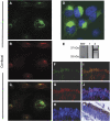
Microarray analysis on pooled samples has previously identified ZDHHC9 (DHHC9) to be upregulated in colon adenocarcinoma compared to normal colon mucosa. Analyses of 168 samples from proximal and distal adenocarcinomas using U133plus2.0 microarrays validated these findings, showing a significant two-fold (log 2) upregulation of DHHC9 transcript (P<10(-6)). The upregulation was more striking in microsatellite stable (MSS), than in microsatellite instable (MSI), tumours. Genes known to interact with DHHC9 as H-Ras or N-Ras did not show expression differences between MSS and MSI. Immunohistochemical analysis was performed on 60 colon adenocarcinomas, previously analysed on microarrays, as well as on tissue microarrays with 40 stage I-IV tumours and 46 tumours from different organ sites. DHHC9 protein was strongly expressed in MSS compared to MSI tumours, readily detectable in premalignant lesions, compared to the rare expression seen in normal mucosa. DHHC9 was specific for tumours of the gastrointestinal tract and localised to the Golgi apparatus, in vitro and in vivo. Overexpression of DHHC9 decreased the proliferation of SW480 and CaCo2 MSS cell lines significantly. In conclusion, DHHC9 is a gastrointestinal-related protein highly expressed in MSS colon tumours. The palmitoyl transferase activity, modifying N-Ras and H-Ras, suggests DHHC9 as a target for anticancer drug design.
Figures


References
-
- Andersen CL, Jensen JL, Orntoft TF (2004) Normalization of real-time quantitative reverse transcription–PCR data: a model-based variance estimation approach to identify genes suited for normalization, applied to bladder and colon cancer data sets. Cancer Res 64: 5245–5250 - PubMed
-
- Andersen CL, Wiuf C, Kruhoffer M, Korsgaard M, Laurberg S, Orntoft TF (2007) Frequent occurrence of uniparental disomy in colorectal cancer. Carcinogenesis 28: 38–48 - PubMed
-
- Benatti P, Gafa R, Barana D, Marino M, Scarselli A, Pedroni M, Maestri I, Guerzoni L, Roncucci L, Menigatti M, Roncari B, Maffei S, Rossi G, Ponti G, Santini A, Losi L, Di GC, Oliani C, Ponz de LM, Lanza G (2005) Microsatellite instability and colorectal cancer prognosis. Clin Cancer Res 11: 8332–8340 - PubMed
-
- Berthiaume LG (2002) Insider information: how palmitoylation of Ras makes it a signaling double agent. Sci STKE 2002: E41 - PubMed
Publication types
MeSH terms
Substances
LinkOut - more resources
Full Text Sources
Other Literature Sources
Medical
Molecular Biology Databases
Research Materials
Miscellaneous

