Review
. 2007 Jun 18;96(12):1788-95.
doi: 10.1038/sj.bjc.6603813. Epub 2007 May 22.

Mechanisms of adverse effects of anti-VEGF therapy for cancer

Affiliations
Review

Mechanisms of adverse effects of anti-VEGF therapy for cancer

T Kamba et al. Br J Cancer. .

Abstract

Advances in understanding the role of vascular endothelial growth factor (VEGF) in normal physiology are giving insight into the basis of adverse effects attributed to the use of VEGF inhibitors in clinical oncology. These effects are typically downstream consequences of suppression of cellular signalling pathways important in the regulation and maintenance of the microvasculature. Downregulation of these pathways in normal organs can lead to vascular disturbances and even regression of blood vessels, which could be intensified by concurrent pathological conditions. These changes are generally manageable and pose less risk than the tumours being treated, but they highlight the properties shared by tumour vessels and the vasculature of normal organs.


Figures

Figure 1
Figure 1
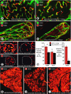
Simple vascular network of tracheal mucosa used to examine effects of VEGF inhibition on normal blood vessels in adult mice. (A) Tracheal vasculature has a simple, repetitive network of arterioles, capillaries, and venules aligned with each cartilaginous ring (Baffert et al, 2004). (BD) Confocal microscopic images of tracheal capillaries showing deposits of fibrin in nonpatent segment of tracheal capillary after inhibition of VEGF signalling by AG-013736 for 1 day. Fibrin deposit (arrow) is shown to be in a nonperfused capillary segment by absence of lectin binding, and is near a region of capillary regression that lacks CD31 immunoreactivity (arrowheads) (Baffert et al, 2006b). (EF) Confocal images of tracheal vasculature showing apoptotic endothelial cells stained for activated caspase-3 (arrow), near region of capillary regression (arrowheads) shown by absence of CD31 immunoreactivity (E). Vascular basement membrane persists after endothelial cells regress, as shown by uninterrupted nidogen immunoreactivity (F) (Baffert et al, 2004). (GH) Confocal micrographs show colocalisation of CD31 (green) and type IV collagen (red) on normal vasculature (G). After AG-013736 for 7 days, empty sleeves of type IV collagen (red, arrows) replace some normal mucosal blood vessels (H) (Inai et al, 2004). Scale bar in (H): 20 μm in (BD); 25 μm in (E) and (F); 30 μm in (G) and (H).
Figure 2
Figure 2
Regression of capillaries in vasculature of normal adult mice after inhibition of VEGF signalling. (AD) Confocal microscopic images showing capillaries in pancreatic islets (A and B) and villi of small intestine (C and D) under baseline conditions and after VEGF inhibition. After Ad-sVEGFR-1 for 14 days, endothelial cells of some capillaries have regressed, leaving pericytes (red, NG2, arrowheads) at sites of regression (Kamba et al, 2006). (E) Comparison of VEGFR-2 and VEGFR-3 immunofluorescence in pancreatic islet capillaries after VEGF inhibition. Stronger endothelial cell VEGFR-2 immunoreactivity under baseline conditions (upper left) than after Ad-sVEGFR-1 for 14 days (upper right). Stronger endothelial cell VEGFR-3 immunoreactivity under baseline conditions (lower left) than after Ad-sVEGFR-1 for 14 days (lower right). (F) Bar graphs showing fluorescence intensities of VEGFR-2 and VEGFR-3 immunoreactivities under baseline conditions and after Ad-sVEGFR-1 for 14 days (Kamba et al, 2006). *P<0.05, significantly different from corresponding control. †P<0.05, significantly different from islets. (GI) Fluorescence micrographs of thyroid capillaries stained for CD31 immunoreactivity show dense vascularity under baseline conditions (G), loss of half of the capillaries after AG-013736 for 7 days (H), and complete regrowth of vasculature during 14 days after end of treatment (I) (Kamba et al, 2006). Scale bar in I: 25 μm in (AD); 40 μm in (E) and (F); 160 μm in (GI).
Figure 3
Figure 3
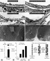
Reduction in endothelial fenestrations (arrowheads) after inhibition of VEGF signalling. (A and B) Transmission electron microscopic images of islet capillaries showing thin endothelium and abundant fenestrations with diaphragms under baseline conditions compared to thick endothelium, few fenestrations, and abundant caveolae after AG-013736 for 21 days (Kamba et al, 2006). (C and D) Transmission EM images of renal glomerular capillaries comparing thin endothelium and abundant fenestrations under baseline conditions with thick endothelium and few fenestrations after Ad-sVEGFR-1 for 14 days (Kamba et al, 2006). (E and F) Scanning electron microscopic images of luminal surface of glomerular capillaries showing abundant endothelial fenestrations under baseline conditions and few fenestrations after Ad-sVEGFR-1 for 14 days (Kamba et al, 2006). (G) Bar graph showing significantly higher concentration of TSH in serum as a measure of altered thyroid function after AG-013736 for 21 days. (H) Bar graph showing increasing amount of proteinuria, indicated by proportion of mice with Albustix values of ++ or greater (⩾100 mg albumin/dl of urine), with increasing dose of AG-013736 for 7 days. (I) Diagram of hypothetical shuttling of diaphragms between endothelial fenestrations and caveolae, with VEGF inhibition driving the process to the right and VEGF signalling driving it to the left (Kamba et al, 2006). Scale bars: 0.3 μm in (A) and (B); 1 μm in (C) and (D); 0.5 μm in (E) and (F).

Similar articles

Cited by

References

    1. Allen JA, Adlakha A, Bergethon PR (2006) Reversible posterior leukoencephalopathy syndrome after bevacizumab/FOLFIRI regimen for metastatic colon cancer. Arch Neurol 63: 1475–1478 - PubMed
    1. Baffert F, Le T, Sennino B, Thurston G, Kuo CJ, Hu-Lowe D, McDonald DM (2006a) Cellular changes in normal blood capillaries undergoing regression after inhibition of VEGF signaling. Am J Physiol Heart Circ Physiol 290: H547–H559 - PubMed
    1. Baffert F, Le T, Thurston G, McDonald DM (2006b) Angiopoietin-1 decreases plasma leakage by reducing number and size of endothelial gaps in venules. Am J Physiol Heart Circ Physiol 290: H107–H118 - PubMed
    1. Baffert F, Thurston G, Rochon-Duck M, Le T, Brekken R, McDonald DM (2004) Age-related changes in vascular endothelial growth factor dependency and angiopoietin-1-induced plasticity of adult blood vessels. Circ Res 94: 984–992 - PubMed
    1. Bates DO, Jones RO (2003) The role of vascular endothelial growth factor in wound healing. Int J Low Extrem Wounds 2: 107–120 - PubMed

Publication types

MeSH terms