Comparative Study
. 2007 May;115(5):734-42.
doi: 10.1289/ehp.9867. Epub 2007 Jan 29.

Molecular mechanisms of the diabetogenic effects of arsenic: inhibition of insulin signaling by arsenite and methylarsonous acid

Affiliations
Comparative Study

Molecular mechanisms of the diabetogenic effects of arsenic: inhibition of insulin signaling by arsenite and methylarsonous acid

David S Paul et al. Environ Health Perspect. 2007 May.

Abstract

Background: Increased prevalences of diabetes mellitus have been reported among individuals chronically exposed to inorganic arsenic (iAs). However, the mechanisms underlying the diabetogenic effects of iAs have not been characterized. We have previously shown that trivalent metabolites of iAs, arsenite (iAs(III)) and methylarsonous acid (MAs(III)) inhibit insulin-stimulated glucose uptake (ISGU) in 3T3-L1 adipocytes by suppressing the insulin-dependent phosphorylation of protein kinase B (PKB/Akt).

Objectives: Our goal was to identify the molecular mechanisms responsible for the suppression of PKB/Akt phosphorylation by iAs(III) and MAs(III).

Methods: The effects of iAs(III) and MAs(III) on components of the insulin-activated signal transduction pathway that regulate PKB/Akt phosphorylation were examined in 3T3-L1 adipocytes.

Results: Subtoxic concentrations of iAs(III) or MAs(III) had little or no effect on the activity of phosphatidylinositol 3-kinase (PI-3K), which synthesizes phosphatidylinositol-3,4,5-triphosphate (PIP(3)), or on phosphorylation of PTEN (phosphatase and tensin homolog deleted on chromosome ten), a PIP(3) phosphatase. Neither iAs(III) nor MAs(III) interfered with the phosphorylation of 3-phosphoinositide-dependent kinase-1 (PDK-1) located downstream from PI-3K. However, PDK-1 activity was inhibited by both iAs(III) and MAs(III). Consistent with these findings, PDK-1-catalyzed phosphorylation of PKB/Akt(Thr308) and PKB/Akt activity were suppressed in exposed cells. In addition, PKB/Akt(Ser473) phosphorylation, which is catalyzed by a putative PDK-2, was also suppressed. Notably, expression of constitutively active PKB/Akt restored the normal ISGU pattern in adipocytes treated with either iAs(III) or MAs(III).

Conclusions: These results suggest that inhibition of the PDK-1/PKB/Akt-mediated transduction step is the key mechanism for the inhibition of ISGU in adipocytes exposed to iAs(III) or MAs(III), and possibly for impaired glucose tolerance associated with human exposures to iAs.


Figures

Figure 1
Figure 1
iAsIII and MAsIII inhibit ISGU by adipocytes at concentrations that do not compromise cell viability. 3T3-L1 adipocytes were treated with iAsIII (A) or MAsIII (B) for 4 hr. [14C]-2-Deoxyglucose uptake was assessed in treated and untreated (control) cells after a 10-min activation with insulin. Cell viability was measured by MTT assay. Each value represents the mean ± SD; n = 3–5 experiments. *Statistically significant differences (p < 0.05) between treated and control cells. The concentrations of 50 μM iAsIII and 2 μM MAsIII (indicated by arrows) were used in all subsequent experiments.
Figure 2
Figure 2
ISGU in adipocytes exposed to subtoxic concentrations of iAsIII and MAsIII is independent of caspase-3 activation. (A) Caspase-3 activity was measured in 3T3-L1 adipocytes treated for 4 hr with 50 μM iAsIII or 2 μM MAsIII and in untreated (control) cells in the presence or absence of 75 μM AC-DEVD-CHO, a caspase-3 inhibitor. Adipocytes treated with 500 μM H2O2 for 4 hr were used as positive controls. (B) Basal and insulin-stimulated uptake of [14C]-2-deoxyglucose was measured in adipocytes exposed for 4 hr to 50 μM iAsIII or 2 μM MAsIII in the presence or absence of 75 μM AC-DEVD-CHO. Each value represents the mean ± SD; n = 3–5 experiments. *Statistically significant differences (p < 0.05) between treated and untreated (control) cells.
Figure 3
Figure 3
Four-hour exposures to subtoxic concentrations of iAsIII or MAsIII do not increase DNA fragmentation in cultured adipocytes. DNA fragmentation was measured by TUNEL in 3T3-L1 adipocytes treated with 50 μM iAsIII or 2 μM MAsIII for 4, 24, 48, and 72 hr. Untreated adipocytes were used as controls. Color images show green fluorescein signal of fragmented DNA in apoptotic cells. Gray-scale images illustrate the corresponding cell morphology. Representative fields of two independent experiments are shown. Bars = 40 μm.
Figure 4
Figure 4
Retention of As species in 3T3-L1 adipocytes exposed to iAsIII or MAsIII. As species retained in 3T3-L1 adipocytes exposed for 4 hr to 50 μM iAsIII or 2 μM MAsIII were analyzed by HG-AAS. Note: The HG-AAS technique used in this study cannot distinguish between AsIII and AsV species. Each value represents the mean ± SD; n = 3 experiments.
Figure 5
Figure 5
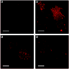
Exposures to subtoxic concentrations of iAsIII or MAsIII inhibit GLUT4 association with the plasma membrane of insulin-activated adipocytes. Immunofluorescent images of GLUT4 in plasma membrane lawns isolated from control (untreated) 3T3-L1 adipocytes before (A) and after activation (B) with insulin and from insulin-activated adipocytes treated for 4 hr with 50 μM iAsIII (C) or 2 μM MAsIII (D). Adipocytes were fixed and sonicated to prepare plasma membrane lawns. GLUT4 was labeled with an anti-GLUT4 antibody and visualized with a fluorescent secondary antibody. Representative fields of two independent experiments are shown. Bars = 10 μm.
Figure 6
Figure 6
Exposures to subtoxic concentrations of iAsIII or MAsIII do not affect insulin signal mediators that regulate PIP3 levels in insulin-activated adipocytes. (A) Immunoblot analyses of the activated PI-3K, total PTEN, and phosphorylated PTEN (Ser380) in control 3T3-L1 adipocytes before or after activation with insulin and in insulin-activated adipocytes treated for 4 hr with 50 μM iAsIII or 2 μM MAsIII. Activated PI-3K was immunoprecipitated from control and exposed cells with an anti-phospho-Tyr (PY20) antibody and immunoblotted with an antibody against the regulatory (p85) subunit. Representative blots of three independent experiments are shown. (B) The ratio of phosphorylated PTEN (Ser380) to total PTEN expressed as a percent of the ratio found in control adipocytes before activation with insulin. Each value represents the mean ± SD; n = 3 experiments.
Figure 7
Figure 7
Exposures to subtoxic concentrations of AsIII or MAsIII do not affect the phosphorylation of PDK-1 but inhibit PDK-1 activity in adipocytes. (A) Immunoblot analysis of Ser241-phosphorylated PDK-1 in insulin-activated 3T3-L1 adipocytes treated for 4 hr with 50 μM iAsIII or 2 μM MAsIII and in control (untreated) insulin-activated adipocytes. Representative blot of three independent experiments is shown. (B) The kinase activity of PDK-1 immunoprecipitated from insulin-activated control or treated 3T3-L1 adipocytes. Each value represents the mean ± SD; n = 4–5 experiments. *Statistically significant differences (p < 0.05) between treated and control cells.
Figure 8
Figure 8
Exposures to subtoxic concentrations of iAsIII or MAsIII inhibit the phosphorylation and PKB/Akt. (A) Immunoblot analysis of total PKB/Akt and Ser473- or Thr308-phosphorylated PKB/Akt in insulin-activated 3T3-L1 adipocytes treated for 4 hr with 50 μM iAsIII or 2 μM MAsIII and in insulin-activated control (untreated) cells. Representative blots of three independent experiments are shown. (B) The activity of PKB/Akt immunoprecipitated from insulin-activated control and treated adipocytes. Each value represents the mean ± SD; n = 4–5 experiments. *Statistically significant differences (p < 0.05) between treated and control cells.
Figure 9
Figure 9
Constitutive activation of protein kinase B (PKB/Akt) prevents the inhibition of insulin-stimulated glucose uptake in adipocytes treated with subtoxic concentrations of iAsIII or MAsIII. Basal and insulin-stimulated [14C]-2-deoxyglucose uptake by 3T3-L1 adipocytes treated for 4 hr with 50 μM iAsIII or 2 μM MAsIII and by untreated adipocytes expressing a constitutively active myr-PKB/Akt) (black bar, an inactive PKB/Akt mutant (A2myr-PKB/Akt) (blue bar), or an empty expression vector (white bar). Each value represents the mean ± SD; n = 4–5 experiments. *Statistically significant differences (p < 0.05) between treated and control cells.
Figure 10
Figure 10
The molecular mechanism underlying the inhibition of insulin-stimulated glucose uptake by adipocytes exposed to iAsIII or MAsIII. The inhibition of PDK-1 and putative PDK-2 activities by iAsIII and MAsIII results in suppression of the downstream signaling steps, including PKB/Akt phosphorylation and GLUT4 translocation to the plasma membrane.

Similar articles

Cited by

References

    1. Alessi DR, Deak M, Casamayor A, Caudwell FB, Morrice N, Norman DG, et al. 3-Phosphoinositide-dependent protein kinase-1 (PDK1): structural and functional homology with the Drosophila DSTPK61 kinase. Curr Biol. 1997;7:776–789. - PubMed
    1. Altamirano MM, Libreros-Minotta CA, Lara-Lemus R, Calcagno M. Evidence for vicinal thiols and their functional role in glucosamine-6-phosphate deaminase from Escherichia coli. Arch Biochem Biophys. 1989;269:555–561. - PubMed
    1. Augustine JA, Sutor SL, Abraham RT. Interleukin 2- and polyomavirus middle T antigen-induced modification of phosphatidylinositol 3-kinase activity in activated T lymphocytes. Mol Cell Biol. 1991;11:4431–4440. - PMC - PubMed
    1. Bazuine M, Carlotti F, Tafrechi RS, Hoeben RC, Maassen JA. Mitogen-activated protein kinase (MAPK) phosphatase-1 and -4 attenuate p38 MAPK during dexamethasone-induced insulin resistance in 3T3-L1 adipocytes. Mol Endocrinol. 2004;18:1697–1707. - PubMed
    1. Bazuine M, Ouwens DM, Gomes de Mesquita DS, Maassen JA. Arsenite stimulated glucose transport in 3T3-L1 adipocytes involves both Glut4 translocation and p38 MAPK activity. Eur J Biochem. 2003;270:3891–3903. - PubMed

Publication types

MeSH terms