. 2007 May 24;54(4):547-57.
doi: 10.1016/j.neuron.2007.04.029.

Zfp423/OAZ participates in a developmental switch during olfactory neurogenesis

Affiliations

Zfp423/OAZ participates in a developmental switch during olfactory neurogenesis

Li E Cheng et al. Neuron. .

Abstract

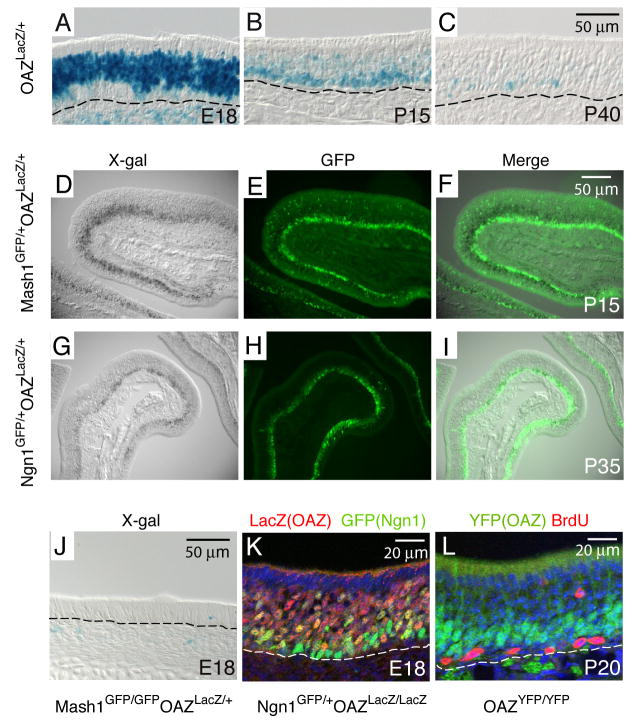
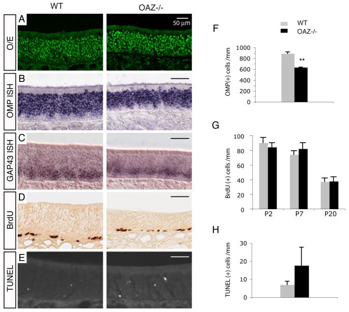
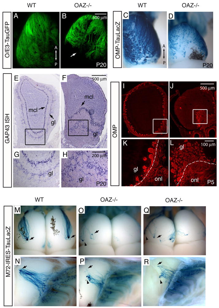
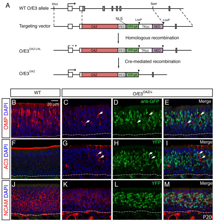
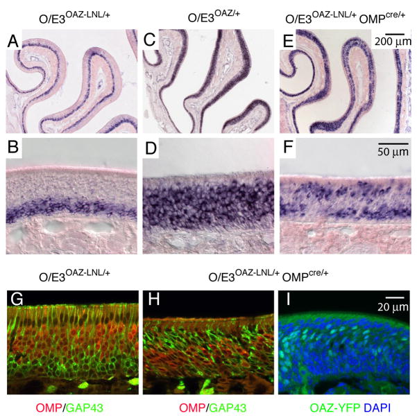
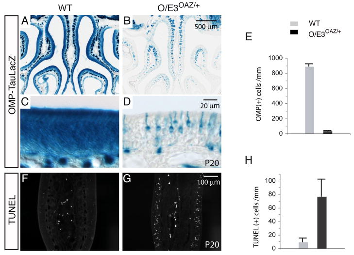
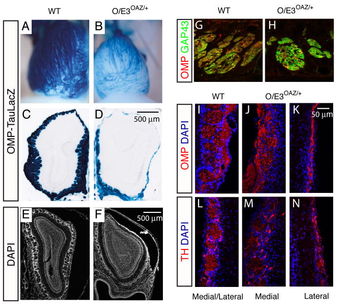
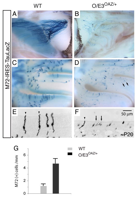
The coordination of gene expression is critical for cell differentiation and the subsequent establishment of tissue function. We show here that a multiple zinc finger transcription factor, Zfp423/OAZ, is transiently expressed in newly differentiating olfactory-receptor neurons (ORNs) and has a key role in coordinating the expression of immature and mature stage-specific genes. OAZ deletion in mice impairs aspects of ORN differentiation, particularly the patterns of axonal projection to the olfactory bulb. OAZ gain-of-function experiments show that sustained OAZ expression throughout ORN maturation arrests ORN development at an immature stage and alters OR gene expression. Importantly, reintroducing OAZ expression in mature ORNs suppresses mature marker expression and reactivates immature-specific markers. Together, these experiments suggest that OAZ participates in a developmental switch regulating the transition from differentiation to maturation in ORNs.


Figures

Figure 1
Figure 1. OAZ is expressed in newly differentiating ORNs
(A) In OAZlacZ/+ mice, X-gal positive cells are present in a broad region in the OE at E18. (B, C) At P15 and P40, X-gal positive cells become restricted to the basal OE. The basal lamina is indicated by dashed line. (D-F) OAZ is expressed downstream of Mash1. OAZ-expressing cells were identified by X-gal staining (D) and Mash1-expressing cells were identified with an anti-GFP antibody (E). In the merged picture (F), X-gal positive cells are located above GFP-positive cells. (G-I) In OAZlacZ/+Ngn1GFP/+ mice, X-gal positive OAZ-expressing cells are in the same layer as GFP-positive Ngn1-expressing cells. (J) OAZ-expressing cells are absent in Mash1-null mice. (K) Colocalization of OAZ and Ngn1 in the OE of OAZlacZ/lacZNgn1GFP/+ mice. (L) OAZ is expressed in postmitotic cells in the OE. OAZ-expressing cells were identified in homozygous OAZYFP/YFP mice (see figure 2) with an anti-GFP antibody (green) and proliferating cells were labeled with BrdU (red).
Figure 2
Figure 2. Characterization of OE in OAZ−/− mice
(A–E) O/E immunostaining, OMP and GAP43 in situ hybridization, BrdU labeling and TUNEL staining in wild-type and OAZ−/− mice. There is a reduction of mature cells and a two-fold increase of apoptotic cells in OAZ−/− mice. (F) Quantification of OMP-positive cells reveals a 30% reduction in OAZ−/− mice (wt, 890±30/mm; OAZ−/−, 635±15/mm; n=3, p<0.001). (G) Quantification of BrdU-positive cells in the OE at P2, P7 and P20 (n=2 mice for each genotype at each age group). (H) Quantification of TUNEL-positive cells in the OE (wt, 7.0±1.9/mm, n=6; OAZ−/−, 17.6±10/mm, n=5, p<0.03). Mice examined were from P20-P25 except that BrdU labeling was conducted at three ages.
Figure 3
Figure 3. OAZ−/− mice exhibit ORN projection defects
(A–D) The ORN projection to the OB is visualized by O/E3-tauGFP reporter (A, B) and OMP-taulacZ reporter (C, D). In whole-mount dorsal view, ORN axons fail to innervate the caudal OB in OAZ−/− mice (n>6). (E, H) GAP43 in situ hybridization labels mitral cells and periglomerular cells in the OB. In OAZ−/− mice, the dorsal OB surface is devoid of glomeruli. The mitral cell layer (mcl) at the ventral OB is pushed inwardly by the abnormally accumulated glomeruli. (I–L) Labeling of olfactory axons with OMP antibody reveals tangled axons and disorganized glomeruli on the ventral surface in a P5 OAZ−/− mouse. (M–R) Poor convergence and a ventral shift of the M72 glomeruli in OAZ−/− mice. Projection of the M72-expressing axons is visualized by X-gal staining in M72-IRES-taulacZ reporter mice. As M72-expressing neurons converge to one lateral and one medial glomerulus in each bulb in control mice (arrows in M, N), the M72 glomerulus in OAZ−/− mice is shifted ventrally (arrowheads in O-R, n=6) and a subset of axons miss their target. All reporter mice examined were from P20-P25.
Figure 4
Figure 4. Sustained OAZ expression arrests ORN maturation
(A) Schematic representation of the OAZ knock-in strategy. Full length OAZ cDNA, followed by IRES-3NLS-YFP-pA and LTNL cassette was inserted in the 5′UTR of the O/E3 gene, replacing the first five exons of O/E3. Transcription initiation sites are indicated by arrow. The LTNL cassette can be deleted by CRE-mediated recombination. (B-M) OMP, AC3 and NCAM staining in wild-type (B, F, J) and O/E3OAZ/+ mice (C, G, K) at P20 revealed a dramatic reduction of mature cells in O/E3OAZ/+ mice. The distribution of OAZ-expressing cells was visualized by either GFP antibody (D) or direct YFP fluorescence (H, L). The merged images (E, I, M) show that OAZ expression does not overlap with mature markers but overlaps with neuronal marker NCAM.
Figure 5
Figure 5. OAZ expression is sufficient for an immature phenotype
(A–D) GAP43 in situ hybridization demonstrates that the majority of cells in the epithelium are arrested at an immature stage in O/E3OAZ/+ mice and the absolute number of GAP43-positive cells in the epithelium is increased. (E–F) In O/E3OAZ-LNL/+OMPCRE/+ mice, in which OAZ-YFP is selectively expressed in mature cells, a second population of GAP43-expressing cell is identified at the apical epithelium. (G, H) OMP/GAP43 double labeling in O/E3OAZ-LNL/+OMPCRE/+ mice shows that OMP and GAP43 are regulated in a reciprocal fashion by OAZ; in the apical GAP43-expressing cells, OMP expression is repressed. (I) OAZ-YFP reporter shows the reintroduction of OAZ expression (green) in mature cells in O/E3OAZ-LNL/+OMPCRE/+ mice. Mice examined were from P20-P25.
Figure 6
Figure 6. Defects of ORN maturation and increased apoptosis in O/E3OAZ/+ mice
(A–D) X-gal staining of the OMP-taulacZ reporter confirms the reduction of mature cells in O/E3OAZ/+ mice. (E) Quantification of OMP-positive cells in wild-type and O/E3OAZ/+ mice (wt, 865±66/mm; O/E3OAZ/+, 47±28/mm, n=3, P<0.001). (F, G) TUNEL staining shows a dramatic increase of apoptotic cells in the OE of O/E3OAZ/+ mice. (H) Quantification of TUNEL-positive cells (wt, 9.4±5.4/mm, n=3; O/E3OAZ/+, 76.5±25.2/mm, n=3, P<0.01).
Figure 7
Figure 7. Defects in the ORN projection in O/E3OAZ/+ mice
(A, B) Whole-mount X-gal staining reveals that the dorsal OB is largely devoid of glomeruli. (C–F) Coronal sections through the OB confirm the reduced ONL and absence of dorsal glomeruli in O/E3OAZ/+ mice. (G, H) OMP/GAP43 double-labeling of axon bundles. In O/E3OAZ/+ mice, axon bundles are GAP43-positive and a small fraction of axons are OMP-positive, suggesting that GAP43-expressing immature cells initiate their axonal growth in O/E3OAZ/+ mice. (I–K) OMP staining demonstrates that the medial OB surface contains OMP-positive olfactory axons and disorganized glomerular structures, while the lateral surface is covered by thin ONL. (L–N) The expression of tyrosine hydroxylase in periglomerular interneurons is greatly reduced at the lateral OB in O/E3OAZ/+ mice. Mice examined were from P20-P25.
Figure 8
Figure 8. Sustained OAZ expression alters OR gene expression
(A, B) Whole-mount X-gal staining of M72-IRES-taulacZ reporter. The axons from M72-expressing cells in O/E3OAZ/+ mice do not converge, but project along the dorsal to ventral surface of the OB. (C, D) In the O/E3OAZ/+ epithelium, the strongly stained axon-projecting M72-expressing cells are accompanied by a large number of weakly stained non-projecting cells (arrows). (E, F) In coronal sections, the weakly stained cells (arrows) display short dendrites and no obvious axon extensions. The signal below the basal lamina reflects individual axons stained in cross sections. (G) Quantification of M72-expressing cells in control and O/E3OAZ/+ mice (wt, 1.2±0.3/mm, n=3; O/E3OAZ/+, 4.7±0.7/mm, n=3, P<0.002). Mice examined were from P20-P25.

Similar articles

Cited by

References

    1. Alcaraz WA, Gold DA, Raponi E, Gent PM, Concepcion D, Hamilton BA. Zfp423 controls proliferation and differentiation of neural precursors in cerebellar vermis formation. Proc Natl Acad Sci U S A 2006 - PMC - PubMed
    1. Bakalyar HA, Reed RR. Identification of a specialized adenylyl cyclase that may mediate odorant detection. Science. 1990;250:1403–1406. - PubMed
    1. Baker H, Cummings DM, Munger SD, Margolis JW, Franzen L, Reed RR, Margolis FL. Targeted deletion of a cyclic nucleotide-gated channel subunit (OCNC1): biochemical and morphological consequences in adult mice. J Neurosci. 1999;19:9313–9321. - PMC - PubMed
    1. Brunet LJ, Gold GH, Ngai J. General anosmia caused by a targeted disruption of the mouse olfactory cyclic nucleotide-gated cation channel. Neuron. 1996;17:681–693. - PubMed
    1. Calof AL, Chikaraishi DM. Analysis of neurogenesis in a mammalian neuroepithelium: proliferation and differentiation of an olfactory neuron precursor in vitro. Neuron. 1989;3:115–127. - PubMed

Publication types

MeSH terms