Review
. 2007 Sep;28(9):1833-46.
doi: 10.1016/j.peptides.2007.04.008. Epub 2007 Apr 20.

Tuning immune tolerance with vasoactive intestinal peptide: a new therapeutic approach for immune disorders

Affiliations
Review

Tuning immune tolerance with vasoactive intestinal peptide: a new therapeutic approach for immune disorders

David Pozo et al. Peptides. 2007 Sep.

Abstract

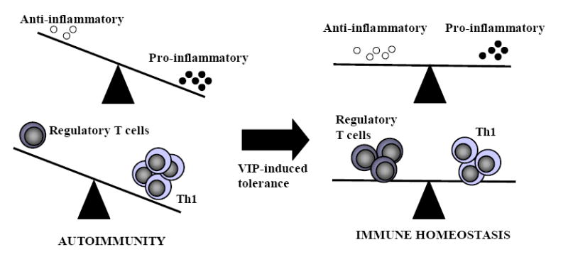
The induction of immune tolerance is essential for the maintenance of immune homeostasis and to limit the occurrence of exacerbated inflammatory and autoimmune conditions. Multiple mechanisms act together to ensure self-tolerance, including central clonal deletion, cytokine deviation and induction of regulatory T cells. Identifying the factors that regulate these processes is crucial for the development of new therapies of autoimmune diseases and transplantation. The vasoactive intestinal peptide (VIP) is a well-characterized endogenous anti-inflammatory neuropeptide with therapeutic potential for a variety of immune disorders. Here, we examine the latest research findings, which indicate that VIP participates in maintaining immune tolerance in two distinct ways: by regulating the balance between pro-inflammatory and anti-inflammatory factors, and by inducing the emergence of regulatory T cells with suppressive activity against autoreactive T-cell effectors.


Figures

Figure 1
Figure 1. VIP restores tolerance in autoimmune disorders by acting at multiple levels
Loss of immune tolerance compromises immune homeostasis and results in the onset of autoimmune disorders (Crohn’s disease is shown as an example). The initial stages of inflammatory bowel disease involve multiple steps that can be divided into two main phases: early events associated with initiation and establishment of autoimmunity to components of the colonic mucosa, and later events associated with the evolving immune and destructive inflammatory responses. Progression of the autoimmune response involves the development of self-reactive T helper 1 (TH1) cells in Peyer’s patches and mesenteric lymph nodes, their entry into the colonic mucosa, release of proinflammatory cytokines (tumor-necrosis factor-α (TNFα) and interferon-γ (IFNγ)) and chemokines and subsequent recruitment and activation of inflammatory cells (macrophages and neutrophils). Inflammatory mediators, such as cytokines, nitric oxide (NO) and free radicals, which are produced by infiltrating cells and resident lamina propria cells, have a crucial role in the destruction of the intestinal epithelium and mucosa. In addition, the TH1-mediated production of IgG2 autoantibodies, which activate complement and neutrophils, contributes to autoimmune pathology. Regulatory T cells are key players in maintaining tolerance by their suppression of self-reactive TH1 cells through a mechanism that involves production of interleukin-10 (IL-10) and transforming growth factor-β (TGFβ), and/or expression of the cytotoxic T-lymphocyte associated antigen 4 (CTLA4). VIP induces immune tolerance and inhibits the autoimmune response through several non-excluding mechanisms. A) VIP decreases TH1-cell functions directly on differentiating T cells, or indirectly via dendritic cell (DC). As a consequence, inflammatory and autoimmune responses are impaired because of reduced infiltration/activation of neutrophils and macrophages by IFNγ and the abolition of the production of complement-fixing IgG2a antibodies. B) VIP inhibits the production of inflammatory cytokines, chemokines and free radicals by macrophages and resident cells. In addition, it impairs the costimulatory activity of macrophages on effector T cells, inhibiting subsequent clonal expansion. This avoids the inflammatory response and its cytotoxic effects against intestinal mucosa and epithelium. C) VIP induces the new generation of peripheral Treg cells that suppress autoreactive T cells activation through a mechanism that involves the production of interleukin-10 (IL-10) and transforming growth factor-β (TGFβ), and/or expression of the cytotoxic T-lymphocyte associated antigen 4 (CTLA4). In addition, VIP indirectly generates Treg cells through the differentiation of tolerogenic DCs. Arrows indicate a stimulatory effect. Back-crossed lines indicate an inhibitory effect.
Figure 2
Figure 2. Molecular mechanisms and transcription factors involved in the anti-inflammatory effects of VIP
The binding of an inflammatory stimulus, such as the bacterial endotoxin lipopolysaccharide (LPS), to the membrane-bound CD14-toll like receptor (TLR) complex in inflammatory cells (i.e., macrophage, microglia and dendritic cells) results in the stimulation of two different pathways involved in the transcription activation of several inflammatory mediators: nuclear factor-κB (NFκB) and a mitogen-activated protein kinase (MAPK) cascade. In unstimulated cells, NFκB is sequestered in the cytosol by its inhibitor IκB. Cellular stimulation results in IκB phosphorylation by a specific kinase (IKK), triggering IκB ubiquitination, and proteosomal degradation, releasing NFκB to translocate to the nucleus where it binds to specific κB promoter elements. Interaction between NFκB and coactivators such as the cAMP-response element binding protein (CREB)-binding protein (CBP) is required for maximal transactivation. Such coactivators bridge various transcriptional activators and components of the basal transcriptional machinery. Meanwhile, the activation of MAPK kinase kinase 1 (MEKK1) activates different MAPKs, leading to the phosphorylation/activation of cJun by Jun kinase (JNK), of TATA-box-binding protein (TBP) by p38MAPK, and of Elk1 by extracellular signal-regulated kinase (ERK1/2). cJun, which together with cFos comprise the activator protein-1 (AP1), which acts to transactivate various inflammatory genes through its binding to AP1 sites and cAMP-response elements (CRE). TBP participates in the initiation of transcription by recruiting the basal transcriptional machinery and various transcription factors through its binding to TATA-box sequences. On the other hand, the binding of IFNγ to its receptor initiates the phosphorylation/activation of the Janus kinases Jak1/Jak2, resulting in the generation of the phosphorylated signal transducer and activator of transcription-1 (STAT1) dimers, their translocation to the nucleus and the expression of the IFN regulatory factor-1 (IRF-1) which in turn transactivates multiple effector genes. The binding of VIP to VPAC1 increases cyclic AMP (cAMP), activates protein kinase A (PKA) and inhibits IKK, stabilizing the inhibitor IκB and preventing nuclear translocation of NFκB p50/p65 complex (1). In addition, PKA activation induces the phosphorylation of CREB which, due to its high affinity for the coactivator CBP, prevents the association of CBP with p65 (2). Furthermore, PKA activation inhibits MEKK1 activation, and the subsequent activation of p38MAPK and TBP (3). Non-phosphorylated TBP lacks the ability to bind to the TATA box, and to form an active transactivating complex with CBP and NFκB, resulting in inefficient recruitment of the RNA polymerase II, which further weakens transcription. Inhibition of MEKK1 also deactivates JNK and cJun phosphorylation (4). In addition, PKA induces the expression of JunB, which can inactivate the transcriptional AP1 complex via displacement of of c-Jun with JunB or CREB. PKA activation can also inhibit both NFκB and MAPK pathways by downregulating TLR and CD14 expression (5). Finally, PKA signaling inhibits Jak-STAT1 pathway and subsequent induction of IRF1 (6). The end result is that the complex of transcriptional transactivators, which were recruited to the promoters of several inflammatory mediators (tumor-necrosis factor-α (TNFα) is shown as an example) in response to the signaling via the TLR4 receptor, is significantly disrupted by neuropeptide treatment (compare profile A with profile B). Arrows indicate a stimulatory effect. Back-crossed lines indicate an inhibitory effect.
Figure 3
Figure 3. VIP generates various populations of regulatory T cells involved in immune tolerance
Immune tolerance depends on the generation of both natural and inducible populations of regulatory T (Treg) cells, which have complementary and overlapping functions in the control of immune responses in vivo. Natural Treg cells develop and migrate from the thymus and constitute 5-10% of peripheral T cells in normal mice and humans. These CD4+CD25+ Treg cells express the transcriptional repressor FoxP3 (forkhead box P3) and cytotoxic T-lymphocyte associated antigen 4 (CTLA4). Natural Treg cells suppress clonal expansion of self-reactive T cells through a mechanism that is cell-cell contact dependent and mediated by CTLA4. Interaction of CTLA4 with CD80 and/or CD86 on the surface of the antigen-presenting cells (APCs) delivers a negative signal for T-cell activation. In vivo studies, but not most in vitro studies, have found a role for cytokines such as IL-10 and transforming growth factor-β (TGFβ) in the function of natural Treg cells. Other populations of antigen-specific Treg cells can be induced from CD4+CD25- or CD8+CD25- T cells in the periphery under the influence of semi-mature tolerogenic dendritic cells and/or various soluble factors such as IL-10, TGFβ1 and interferon-α. The inducible Treg populations consist of distinct subsets: T regulatory 1 (Tr1) cells, which secrete high levels of IL-10 and probably TGFβ1; T helper 3 (Th3) cells, which secrete high levels of TGFβ1; and CD8+ Treg cells, which secrete IL-10. These immunosuppressive cytokines inhibit the proliferation of effector T cells and their production of cytokines, as well as the cytotoxic (CTL) activity of CD8+ T cells, either directly or through their inhibitory action on the maturation/activation of APCs. In addition, CD8+ Treg cells induce the expression of the immunoglobulin-like transcripts ILT3 and ILT4 in APCs, which negatively affect APC function. These suppressive responses can be beneficial for the restoration of immune homeostasis in the host. In treating autoimmune disease, Treg cells suppress autoreactive TH1 responses involved in the destruction of the target tissue. While in transplantation, Treg cells inhibit host CD4+ and CD8+ T cells that can otherwise recognize and react to alloantigens causing transplant rejection. In the case of bone-marrow transplantation Treg cells suppress alloreactive T cells present in the graft that are responsible for causing acute graft-versus-host disease. However, it can also be detrimental as Treg cells can impair effective immune responses to infections, pathogens and tumors. VIP induces the generation of different types of Treg cells by two independent mechanisms. A. The presence of VIP during the initial stages of differentiation of dendritic cells (DCs) from bone-marrow cells or monocytes generates semi-mature DCs that are unable to mature even after adequate activation. Such semi-mature DCs show a tolerogenic phenotype that is characterized by low expression of the co-stimulatory molecules CD40, CD80 and CD86, low production of inflammatory cytokines, such as tumor necrosis factor-α (TNFα), IL-12 and IL-6, and increased secretion of IL-10. It is the lack of essential costimulatory signals and the immunosuppressive cytokine IL-10 in allogeneic or antigen-specific VIP-differentiated tolerogenic DCs that permits them to stimulate the generation of CD4+ and CD8+ T regulatory 1 (Tr1)-like cells. The resulting T cells show the characteristic cytokine profile (high IL-10/transforming growth factor-β1 (TGFβ1) production, little interferon-γ (IFNγ), IL-2, IL-4 secretion), antigen-specific suppressive activity on effector T cells and high expression of the suppressive molecule CTLA4 (cytotoxic T-lymphocyte associated antigen 4) B. VIP also triggers the generation of peripherally induced FoxP3+CD4+CD25+ Treg cells from naïve CD4+CD25- T cells. These cells express high levels of CTLA4 and produce IL-10 and/or TGFβ1. Both VIP-induced Treg cell subtypes contribute to the suppression of self-reactive TH1 cells in autoimmune conditions and alloantigen-specific T cells in transplantation. This can help to restore immune tolerance, and inhibit autoimmunity, transplant rejection and graft-versus-host disease.
Figure 4
Figure 4. VIP restores immune tolerance
An imbalance between regulatory T cells versus TH1 cells, or of anti-inflammatory cytokines versus proinflammatory factors, are key causes of autoimmune disorders. VIP restores immune homeostasis by rebalancing this scenario by downregulating the inflammatory response and inducing regulatory T cells.

Similar articles

Cited by

References

    1. Abad C, Martinez C, Juarranz MG, Arranz A, Leceta J, Delgado M, et al. Therapeutic effects of vasoactive intestinal peptide in the trinitrobenzene sulfonic acid mice model of Crohn’s disease. Gastroenterology. 2003;124:961–71. - PubMed
    1. Bangale Y, Karle S, Planque S, Zhou YX, Taguchi H, Nishiyama Y, et al. VIPase autoantibodies in Fas-defective mice and patients with autoimmune disease. FASEB J. 2003;17:628–35. - PubMed
    1. Banner KH, Trevethick MA. PDE4 inhibition: a novel approach for the treatment of inflammatory bowel disease. Trends Pharmacol Sci. 2004;25:430–6. - PubMed
    1. Blalock JE. The immune system as the sixth sense. J Intern Med. 2005;257:126–38. - PubMed
    1. Bluestone JA. Regulatory T-cell therapy: is it ready for the clinic? Nat Rev Immunol. 2005;5:343–9. - PubMed

Publication types

MeSH terms

Substances