Regulation of cell junction dynamics by cytokines in the testis: a molecular and biochemical perspective
- PMID: 17521954
- PMCID: PMC2701191
- DOI: 10.1016/j.cytogfr.2007.04.009
Regulation of cell junction dynamics by cytokines in the testis: a molecular and biochemical perspective
Abstract
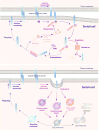
Studies in the past decade in the field have demonstrated the significance of cytokines in regulating epithelial and endothelial cell junctions including tight and anchoring junctions in multiple organs including the testis. There are mounting evidences in recent years that cytokines play a crucial role in the restructuring of junctions at the Sertoli-Sertoli and Sertoli-germ cell interface in the seminiferous epithelium during spermatogenesis. These earlier studies, however, were focused on the effects of cytokines in maintaining the steady-state protein levels of integral membrane proteins at the sites of the blood-testis barrier (BTB) and anchoring junctions at the Sertoli-Sertoli and Sertoli-germ cell interface, such as basal and apical ectoplasmic specialization, respectively. The molecular pathway(s) and/or mechanism(s) underlying these effects remained virtually unexplored until very recently. Herein, we summarize and provide some discussions on studies that focused on the role of cytokines in regulating junction restructuring events in epithelia from a molecular and biochemical perspective. Specifically, we use the adult rat or mouse testis as a model to highlight the significance of transcriptional and translational regulation. Specific areas of research that require further attentions are also highlighted.
Figures


Similar articles
-
Disruption of Sertoli-germ cell adhesion function in the seminiferous epithelium of the rat testis can be limited to adherens junctions without affecting the blood-testis barrier integrity: an in vivo study using an androgen suppression model.J Cell Physiol. 2005 Oct;205(1):141-57. doi: 10.1002/jcp.20377. J Cell Physiol. 2005. PMID: 15880438
-
Regulation of Sertoli-germ cell adherens junction dynamics via changes in protein-protein interactions of the N-cadherin-beta-catenin protein complex which are possibly mediated by c-Src and myotubularin-related protein 2: an in vivo study using an androgen suppression model.Endocrinology. 2005 Mar;146(3):1268-84. doi: 10.1210/en.2004-1194. Epub 2004 Dec 9. Endocrinology. 2005. PMID: 15591133
-
c-Yes regulates cell adhesion at the blood-testis barrier and the apical ectoplasmic specialization in the seminiferous epithelium of rat testes.Int J Biochem Cell Biol. 2011 Apr;43(4):651-65. doi: 10.1016/j.biocel.2011.01.008. Epub 2011 Jan 21. Int J Biochem Cell Biol. 2011. PMID: 21256972 Free PMC article.
-
Cytokines and junction restructuring events during spermatogenesis in the testis: an emerging concept of regulation.Cytokine Growth Factor Rev. 2009 Aug;20(4):329-38. doi: 10.1016/j.cytogfr.2009.07.007. Epub 2009 Aug 3. Cytokine Growth Factor Rev. 2009. PMID: 19651533 Free PMC article. Review.
-
Extracellular matrix: recent advances on its role in junction dynamics in the seminiferous epithelium during spermatogenesis.Biol Reprod. 2004 Aug;71(2):375-91. doi: 10.1095/biolreprod.104.028225. Epub 2004 Apr 28. Biol Reprod. 2004. PMID: 15115723 Review.
Cited by
-
The Drosophila cyst stem cell lineage: Partners behind the scenes?Spermatogenesis. 2012 Jul 1;2(3):145-157. doi: 10.4161/spmg.21380. Spermatogenesis. 2012. PMID: 23087834 Free PMC article.
-
Fascin - An actin binding and bundling protein in the testis and its role in ectoplasmic specialization dynamics.Spermatogenesis. 2015 Feb 23;5(1):e1002733. doi: 10.1080/21565562.2014.1002733. eCollection 2015 Jan-Apr. Spermatogenesis. 2015. PMID: 26413410 Free PMC article.
-
TGF-beta3 and TNFalpha perturb blood-testis barrier (BTB) dynamics by accelerating the clathrin-mediated endocytosis of integral membrane proteins: a new concept of BTB regulation during spermatogenesis.Dev Biol. 2009 Mar 1;327(1):48-61. doi: 10.1016/j.ydbio.2008.11.028. Epub 2008 Dec 7. Dev Biol. 2009. PMID: 19103189 Free PMC article.
-
Downstream genes of Sox8 that would affect adult male fertility.Sex Dev. 2009;3(1):16-25. doi: 10.1159/000200078. Epub 2009 Apr 1. Sex Dev. 2009. PMID: 19339814 Free PMC article.
-
Contribution and Regulation of HIF-1α in Testicular Injury Induced by Diabetes Mellitus.Biomolecules. 2025 Aug 19;15(8):1190. doi: 10.3390/biom15081190. Biomolecules. 2025. PMID: 40867634 Free PMC article. Review.
References
-
- de Kretser DM, Kerr JB. The cytology of the testis. In: Knobil E, Neill J, editors. The Physiology of Reproduction. 1. Vol. 837. New York: Raven Press; pp. 932–1988.
-
- Parvinen M. Regulation of the seminiferous epithelium. Endocr Rev. 1982;3:404–17. - PubMed
-
- Hess RA, Schaeffer DJ, Eroschenko VP, Keen JE. Frequency of the stages in the cycle of the seminiferous epithelium in the rat. Biol Reprod. 1990;43:417–524. - PubMed
-
- Dym D. Basement membrane regulation of Sertoli cells. Endocr Rev. 1994;15:102–15. - PubMed
-
- Weber JE, Russell LD, Wong V, Peterson RN. Three-dimensional reconstruction of a rat stage V Sertoli cell. II. Morphometry of Sertoli-Sertoli and Sertoli-germ cell relationships. Am J Anat. 1983;167:163–79. - PubMed
Publication types
MeSH terms
Substances
Grants and funding
LinkOut - more resources
Full Text Sources
Miscellaneous

