. 2007 Jul 20;282(29):21237-43.
doi: 10.1074/jbc.M701341200. Epub 2007 May 22.

Atherogenic phospholipids attenuate osteogenic signaling by BMP-2 and parathyroid hormone in osteoblasts

Affiliations

Atherogenic phospholipids attenuate osteogenic signaling by BMP-2 and parathyroid hormone in osteoblasts

Michael S Huang et al. J Biol Chem. .

Abstract

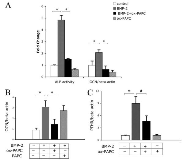
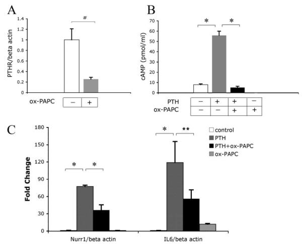
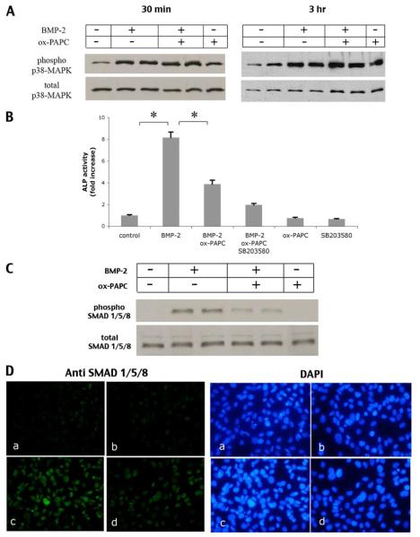
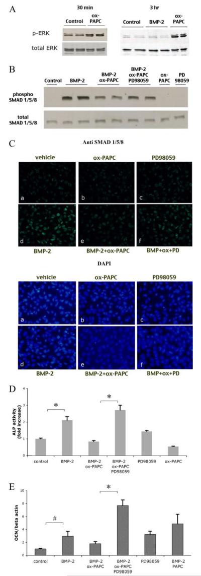
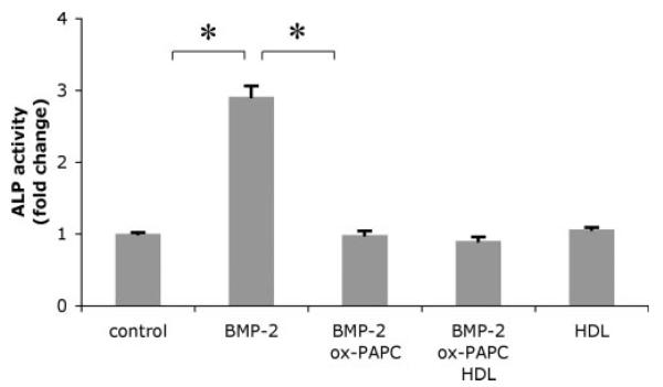
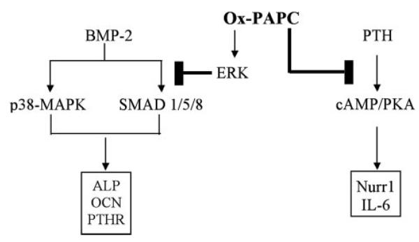
Cardiovascular disease, such as atherosclerosis, has been associated with reduced bone mineral density and fracture risk. A major etiologic factor in atherogenesis is believed to be oxidized phospholipids. We previously found that these phospholipids inhibit spontaneous osteogenic differentiation of marrow stromal cells, suggesting that they may account for the clinical link between atherosclerosis and osteoporosis. Currently, anabolic agents that promote bone formation are increasingly used as a new treatment for osteoporosis. It is not known, however, whether atherogenic phospholipids alter the effects of bone anabolic agents, such as bone morphogenetic protein (BMP)-2 and parathyroid hormone (PTH). Therefore we investigated the effects of oxidized 1-palmitoyl-2-arachidonoyl-sn-glycero-3-phosphorylcholine (ox-PAPC) on osteogenic signaling induced by BMP-2 and PTH in MC3T3-E1 cells. Results showed that ox-PAPC attenuated BMP-2 induction of osteogenic markers alkaline phosphatase and osteocalcin. Ox-PAPC also inhibited both spontaneous and BMP-induced expression of PTH receptor. Consistently, pretreatment of cells with ox-PAPC inhibited PTH-induced cAMP production and expression of immediate early genes Nurr1 and IL-6. Results from immunofluorescence and Western blot analyses showed that inhibitory effects of ox-PAPC on BMP-2 signaling were associated with inhibition of SMAD 1/5/8 but not p38-MAPK activation. These effects appear to be due to ox-PAPC activation of the ERK pathway, as the ERK inhibitor PD98059 reversed ox-PAPC inhibitory effects on BMP-2-induced alkaline phosphatase activity, osteocalcin expression, and SMAD activation. These results suggest that atherogenic lipids inhibit osteogenic signaling induced by BMP-2 and PTH, raising the possibility that hyperlipidemia and atherogenic phospholipids may interfere with anabolic therapy.


Figures

FIGURE 1
FIGURE 1. Effect of ox-PAPC on BMP-induced osteoblastic differentiation
A, alkaline phosphatase activity and osteocalcin expression in response to BMP-2 and ox-PAPC. For the alkaline phosphatase activity assay, cells were cotreated for 2 days with vehicle control, BMP-2 (50 ng/ml), and/or ox-PAPC (10 μg/ml). For osteocalcin expression, RNA was isolated from cells pretreated with ox-PAPC (10 μg/ml) for 4 days, followed by treatment with ox-PAPC and/or BMP-2 (50 ng/ml) for an additional 4 days. Expression was assessed by real-time RT-PCR and normalized to β-actin. B, osteocalcin expression in response to BMP-2, ox-PAPC, and native PAPC. RNA was isolated from cells treated for 3 days with BMP-2 (50 ng/ml) along with either ox-PAPC (10 μg/ml) or PAPC (10 μg/ml). Expression was assessed by real-time RT-PCR and normalized to β-actin. C, PTH receptor (PTHR) expression in response to BMP-2 and ox-PAPC. RNA was isolated from cells as in panel A. Expression was assessed by real-time RT-PCR and normalized to β-actin. Results are shown as mean ± S.E. from all experiments. *, p < 0.0001; #, p < 0.005.
FIGURE 2
FIGURE 2. Effect of ox-PAPC on PTH signaling
A, PTH receptor expression in response to ox-PAPC. RNA was isolated from cells treated for 6 days with ox-PAPC (10 μg/ml). Expression was assessed by real-time RT-PCR and normalized to β-actin. B, cAMP production in response to PTH and ox-PAPC. cAMP accumulation in the medium of cells pretreated for 7 days with ox-PAPC, followed by treatment for an additional 30 min with vehicle, PTH (10−8 m), and/or ox-PAPC (10 μg/ml). C, Nurr-1 and IL-6 expression in response to PTH and ox-PAPC. RNA isolated from cells pretreated for 6 days with ox-PAPC, followed by treatment for an additional 2 h with vehicle, PTH (10−8 m), and/or ox-PAPC (10 μg/ml). Expression was assessed by real-time RT-PCR and normalized to β-actin. Results are shown as mean ± S.E. from all experiments. *, p < 0.0001; #, p < 0.005; **, p < 0.05.
FIGURE 3
FIGURE 3. Effect of ox-PAPC on p38-MAPK and ERK
A, activation of p38-MAPK in response to BMP-2 and ox-PAPC. Western analysis of phosphorylated p38-MAPK from cultures treated with vehicle, BMP-2 (100 ng/ml), and/or ox-PAPC (10μg/ml) for 30 min (left panel)or3 h(right panel). Total p38-MAPK was used as a loading control. B, alkaline phosphatase activity in response to BMP-2, ox-PAPC, and SB203580. Cells were pretreated for 30 min with SB203580 (10 μm), followed by treatment for 2 days with vehicle, BMP-2 (100 ng/ml), ox-PAPC (10 μg/ml), and/or SB203580 (10 μm) as indicated. *, p < 0.0001. C, activation of SMADs in response to BMP-2 and ox-PAPC. Western analysis of phosphorylated SMAD 1/5/8 in cells treated for 1.5 h with vehicle, BMP-2 (100 ng/ml), ox-PAPC (30 μg/ml), or BMP-2/ox-PAPC. Total SMAD 1/5/8 was used as a loading control. D, immunofluorescent (fluorescein isothiocyanate) staining of phosphorylated SMAD 1/5/8 (green; left panels) in cells treated for 1.5 h with vehicle (a), ox-PAPC (10 μg/ml) (b), BMP-2 (50 ng/ml) (c), or BMP-2 and ox-PAPC (d). 4′,6-Diamidino-2-phenylindole staining (blue; right panels) was used to visualize cell nuclei (magnification ×400).
FIGURE 4
FIGURE 4. Effect of ERK inhibition on ox-PAPC inhibitory effects
A, activation of ERK in response to BMP-2 and ox-PAPC. Western analysis of phosphorylated ERK (p-ERK) from cell cultures treated for 30 min (left panel) or 3 h(right panel) with BMP-2 (50 ng/ml) or ox-PAPC (10 μg/ml). Total ERK was used as a loading control. B, activation of SMADs in response to BMP-2, ox-PAPC, and PD98059. Western analysis of phosphorylated SMAD 1/5/8 in cells treated for 1.5 h with vehicle, BMP-2 (100 ng/ml), ox-PAPC (30 μg/ml), or PD98059 (5 μm), BMP-2/ox-PAPC, or BMP-2/ox-PAPC/PD98059. Total SMAD 1/5/8 was used as a loading control. C, immunofluorescent (fluorescein isothiocyanate) staining of phosphorylated SMAD 1/5/8 (green; upper panels) in cells pretreated for 30 min with PD98059, followed by treatment for an additional 1.5 h with vehicle (a), ox-PAPC (b), PD98059 (c), BMP-2 (d), BMP-2 and ox-PAPC (e), BMP-2, ox-PAPC, and PD98059 (f) in medium supplemented with 0.5% fetal bovine serum. 4 ,6-Diamidino-2-phenylindole staining (blue; lower panels) was used to visualize cell nuclei (magnification ×400). D, alkaline phosphatase activity in response to BMP-2, ox-PAPC, and PD98059. Cells were treated for 2 days with BMP-2 (50 ng/ml), ox-PAPC (10 μg/ml), and/or PD98059 (5 μm) as indicated. E, osteocalcin expression in response to BMP-2, ox-PAPC, and PD98059. Cells were cotreated for 3 days with vehicle, BMP-2 (50 ng/ml), ox-PAPC (10 μg/ml), PAPC (10 μg/ml), or PD98059 (5 μm) as indicated. *, p < 0.0001; #, p < 0.05.
FIGURE 5
FIGURE 5. Effect of antioxidant particle HDL on ox-PAPC inhibitory effects
Alkaline phosphatase activity in response to BMP-2, ox-PAPC, and HDL. Cells were pretreated for 24 h with HDL (100 μg/ml), followed by treatment for an additional 2 days with vehicle, BMP-2 (50 ng/ml), ox-PAPC (10 μg/ml), and/or HDL as indicated. *, p < 0.0001.
FIGURE 6
FIGURE 6. Proposed inhibitory mechanisms of ox-PAPC on BMP-2 and PTH signaling pathways
Ox-PAPC appears to inhibit BMP-2 signaling by attenuating SMAD 1/5/8 activation via ERK and inhibits PTH signaling by attenuating induction of cAMP.

References

    1. Bagger YZ, Tanko LB, Alexandersen P, Qin G, Christiansen C. J. Intern. Med. 2006;259:598–605. - PubMed
    1. Jie KG, Bots ML, Vermeer C, Witteman JC, Grobbee DE. Calcif. Tissue Int. 1996;59:352–356. - PubMed
    1. Barengolts EI, Berman M, Kukreja SC, Kouznetsova T, Lin C, Chomka EV. Calcif. Tissue Int. 1998;62:209–213. - PubMed
    1. Yamaguchi T, Sugimoto T, Yano S, Yamauchi M, Sowa H, Chen Q, Chihara K. Endocr. J. 2002;49:211–217. - PubMed
    1. Orozco P. Eur. J. Epidemiol. 2004;19:1105–1112. - PubMed

Publication types

MeSH terms

Substances