. 2007 Jul;134(13):2521-31.
doi: 10.1242/dev.003855. Epub 2007 May 23.

Multiple dose-dependent roles for Sox2 in the patterning and differentiation of anterior foregut endoderm

Affiliations

Multiple dose-dependent roles for Sox2 in the patterning and differentiation of anterior foregut endoderm

Jianwen Que et al. Development. 2007 Jul.

Abstract

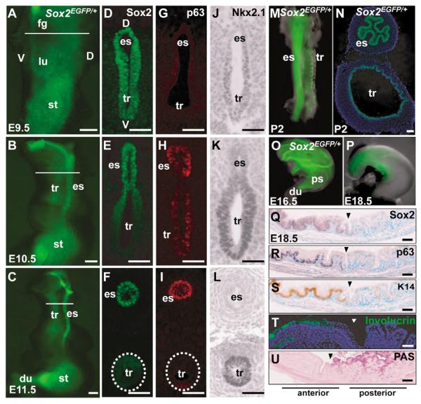
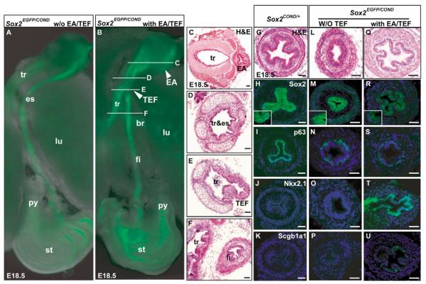
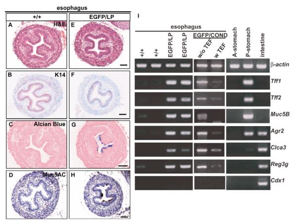
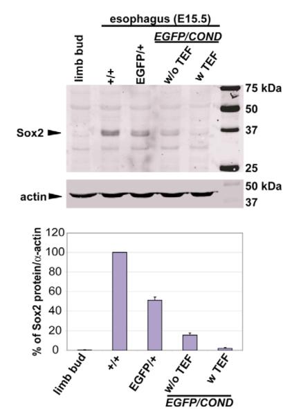
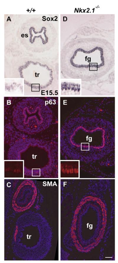
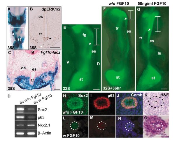
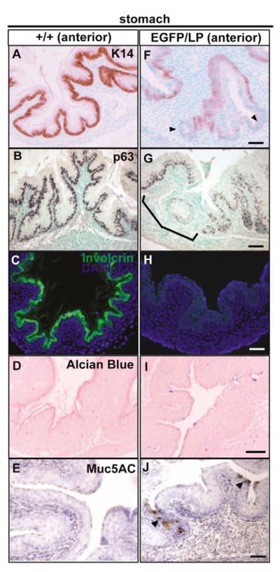
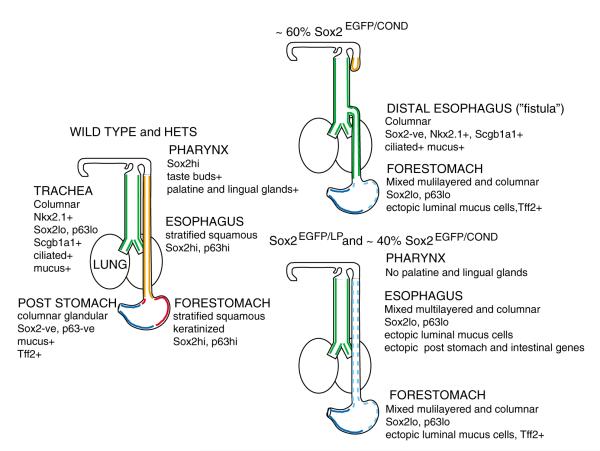
Sox2 is expressed in developing foregut endoderm, with highest levels in the future esophagus and anterior stomach. By contrast, Nkx2.1 (Titf1) is expressed ventrally, in the future trachea. In humans, heterozygosity for SOX2 is associated with anopthalmia-esophageal-genital syndrome (OMIM 600992), a condition including esophageal atresia (EA) and tracheoesophageal fistula (TEF), in which the trachea and esophagus fail to separate. Mouse embryos heterozygous for the null allele, Sox2(EGFP), appear normal. However, further reductions in Sox2, using Sox2(LP) and Sox2(COND) hypomorphic alleles, result in multiple abnormalities. Approximately 60% of Sox2(EGFP/COND) embryos have EA with distal TEF in which Sox2 is undetectable by immunohistochemistry or western blot. The mutant esophagus morphologically resembles the trachea, with ectopic expression of Nkx2.1, a columnar, ciliated epithelium, and very few p63(+) basal cells. By contrast, the abnormal foregut of Nkx2.1-null embryos expresses elevated Sox2 and p63, suggesting reciprocal regulation of Sox2 and Nkx2.1 during early dorsal/ventral foregut patterning. Organ culture experiments further suggest that FGF signaling from the ventral mesenchyme regulates Sox2 expression in the endoderm. In the 40% Sox2(EGFP/COND) embryos in which Sox2 levels are approximately 18% of wild type there is no TEF. However, the esophagus is still abnormal, with luminal mucus-producing cells, fewer p63(+) cells, and ectopic expression of genes normally expressed in glandular stomach and intestine. In all hypomorphic embryos the forestomach has an abnormal phenotype, with reduced keratinization, ectopic mucus cells and columnar epithelium. These findings suggest that Sox2 plays a second role in establishing the boundary between the keratinized, squamous esophagus/forestomach and glandular hindstomach.


Figures

Fig. 1
Fig. 1. Dynamic expression of Sox2EGFP in foregut-derived organs during mouse development
(A-C,M-P) Fluorescence microscopy of foregut organs from Sox2EGFP/+ embryos. N is a cryosection of M. (D-F) Immunohistochemical localization of Sox2 protein in sections of foregut before and after separation into trachea and esophagus. Nuclei are counterstained with DAPI. (G-I) Immunohistochemical localization of p63. (J-L) Immunohistochemical localization of Nkx2.1. (Q-U) Sections through boundary (arrowhead) between stratified keratinized epithelium of the E18.5 forestomach and glandular epithelium of posterior stomach. Immunohistochemistry localizes Sox2 (Q), p63 (R), Keratin 14 (S) and involucrin (T) in keratinized epithelium whereas Periodic Acid Schifff (PAS) staining marks mucus-producing luminal cells of glandular stomach (U). Scale bars: 500 μm in A-C; 50 μm in all others. du, duodenum; es, esophagus; fg, foregut; lu, lung; st, stomach; tr, trachea.
Fig. 2
Fig. 2. Tracheoesophageal fistula and esophageal phenotype of Sox2EGFP/COND hypomorphic mutants
(A,B) Fluorescence microscopy of dissected foregut of Sox2EGFP/COND mutant without (A) and with (B) distal tracheoesophageal fistula (TEF). In B the arrowhead marked EA shows the blind-ended proximal esophagus known in this condition as esophageal atresia (Brunner and van Bokhoven, 2005). White lines indicate level of sections shown in C-F. (C-F) Histological sections through trachea and esophagus of mutant similar to that shown in B. Note the EA in C, and in E the presence of a continuation (TEF) between the dorsal common tube and the distal esophagus (here called the fistula). (G-K) Sections of Sox2COND/+ esophagus demonstrating cellular organization (G), immunohistochemical localization of Sox2, with high-power inset showing strong nuclear staining of basal cells and background cytoplasmic staining of suprabasal cells (H), high p63 in nuclei of basal cells (I) and absence of expression for Nkx2.1 (J) and Scgb1a1 (K). (L-P) Phenotype of esophagus of typical compound mutant without TEF showing cellular organization (note that scale bar is different from G) (L), low Sox2 nuclear staining that is just above background cytoplasmic staining (inset) (M), reduced number of p63-positive cells compared with control (N) and absence of Nkx2.1 and Scgb1a1 (O,P). (Q-U) Phenotype of esophagus of typical compound mutant with TEF, showing cellular organization with a clear monolayered epithelium (Q), absence of Sox2 nuclear staining (R), very few p63-positive cells (S), strong nuclear staining for Nkx2.1 (T) and scattered cells positive for Scgb1a1 (U). Nuclei in H-U are counterstained with DAPI. Scale bars: 100 μm. br, primary bronchus of lung; fi, fistula or distal esophagus; py, pyloric sphincter; tr&es, undivided foregut tube that represents both trachea and esophagus.
Fig. 3
Fig. 3. Abnormal esophagus phenotype of hypomorphic mutants at P0
Sections of esophagi of wild type (A-D) and Sox2EGFP/LP compound mutants (E-H) stained with haematoxylin and eosin (A,E), anti-Keratin 14 (B,F), Alcian blue (C,G), and anti-Muc5AC (D,H). (I) Semi-quantitative RT-PCR for expression in mutant esophagi of several glandular stomach- and intestine-specific genes. Tff1, Tff2 and Muc5B are normally specifically expressed in posterior (antral/fundic) stomach (P-stomach), not in forestomach (A-stomach). Agr2 is normally expressed in both posterior stomach and intestine. Clca3, Reg3g and Cdx1 are normally expressed in intestine. β-actin is internal control for RT-PCR. Scale bars: 50 μm in A-H.
Fig. 4
Fig. 4. Western blot analysis to compare the levels of Sox2 protein in the E15.5 esophagus of mutants with different genotypes and phenotypes
The example shown is typical of three independent experiments. Control sample is from limb bud mesenchyme that does not express Sox2. Quantification showed that the level of Sox2 in Sox2EGFP/COND mutants without fistula was approximately 18% of wild type (mean of three experiments).
Fig. 5
Fig. 5. Dorsalization of the abnormal foregut of Nkx2.1−/− mutants
Histological sections were taken through the undivided foregut of Nkx2.1 mutant embryos just anterior to the abnormal lung sacs, and through the trachea and esophagus of wild-type embryos at about the same level. (A,B) Immunohistochemical analysis of wild-type sections showing high levels of Sox2 (A) and p63 (B) in basal cells of the esophagus, and lower levels in the trachea (higher magnification of trachea shown in the insets). By contrast, the foregut of Nkx2.1-null mutants shows high levels of Sox2 (D) and p63 (E) throughout the basal layer of the undivided foregut (high magnification in the insets). The ectopic expression of Sox2hi and p63hi was less pronounced in more rostral sections of the undivided foregut (data not shown). Immunohistochemistry for smooth muscle actin shows only a localized region of smooth muscle in the wild-type trachea (C) but a continuous layer around the mutant foregut (F). Scale bars: 100 μm. fg, undivided foregut; SMA, smooth muscle actin.
Fig. 6
Fig. 6. Evidence that Fgf10 plays a role in foregut patterning
(A,C) Wholemount and section of foregut of E10.5 FGF10lacZ embryo after staining for β-galactosidase. (A) Ventral view after dissection of the heart. Section in C shows strong Fgf10 expression in ventral mesenchyme. (B) Ventral epithelium (black arrowhead) and mesenchyme (white arrowhead) are positive for phosphorylated ERK1/2 staining. (E-G) Fluorescence microscopy of foreguts from 32-somite stage Sox2EGFP/+ embryo before (E) and after culture for 36 hours without (F) or with (G) 50 ng/ml Fgf10 in the culture medium. White arrowhead marks the junction of the esophagus with the foregut and the white line marks the undivided proximal foregut. The anterior limit was determined from phase microscopy. Note that in samples incubated with Fgf10 the region of undivided foregut is longer than in control incubated without Fgf. (H-O) Section through the esophagus of samples similar to those shown in F,G, stained with antibodies to Sox2 (H,L), p63 (I,M), merged (J,N) and haematoxlyin and eosin (K,O). (H-K) Cultured without Fgf10, (L-O) cultured with Fgf10. The dotted line in K-O outlines the esophageal epithelial layer. Note the difference in length of the scale bars. (D) RT-PCR of transcript levels in isolated esophagi from E11 embryos cultured with and without Fgf10. Primers for p63 were for the transactivating isoform. Scale bars: 100 μm in E-G; 50 μm in H-O.
Fig. 7
Fig. 7. Abnormal phenotype of forestomach of Sox2EGFP/LP mutants
Sections of the wild type (A-E) and mutant (F-J) P0 anterior stomach were stained with anti-Keratin14 (A,F), anti-p63 (B,G), anti-involucrin (C,H), Alcian blue (D,I) and Muc5AC (E,J). Note that Keratin14 expression is reduced or absent (arrowheads) in the mutant forestomach (F). Likewise, anti-p63 immunostaining shows regions of reduced or absent staining in the mutant forestomach (G). Ectopic Alcian blue-positive (I) and Muc5AC-positive (J, arrowheads) cells are present in the mutant forestomach. Scale bars: 50 μm.
Fig. 8
Fig. 8. Summary of the various phenotypes seen in Sox2 hypomorphic mutants
For details, see text.

References

    1. Aubin J, Dery U, Lemieux M, Chailler P, Jeannotte L. Stomach regional specification requires Hoxa5-driven mesenchymal-epithelial signaling. Development. 2002;129:4075–4087. - PubMed
    1. Avilion AA, Nicolis SK, Pevny LH, Perez L, Vivian N, Lovell-Badge R. Multipotent cell lineages in early mouse development depend on SOX2 function. Genes Dev. 2003;17:126–140. - PMC - PubMed
    1. Boyer LA, Lee TI, Cole MF, Johnstone SE, Levine SS, Zucker JP, Guenther MG, Kumar RM, Murray HL, Jenner RG, et al. Core transcriptional regulatory circuitry in human embryonic stem cells. Cell. 2005;122:947–956. - PMC - PubMed
    1. Brunner HG, van Bokhoven H. Genetic players in esophageal atresia and tracheoesophageal fistula. Curr. Opin. Genet. Dev. 2005;15:341–347. - PubMed
    1. Calmont A, Wandzioch E, Tremblay KD, Minowada G, Kaestner KH, Martin GR, Zaret KS. An FGF response pathway that mediates hepatic gene induction in embryonic endoderm cells. Dev. Cell. 2006;11:339–348. - PubMed

Publication types

MeSH terms