. 2007 Aug;81(15):8041-9.
doi: 10.1128/JVI.00068-07. Epub 2007 May 23.

CCR5Delta32 protein expression and stability are critical for resistance to human immunodeficiency virus type 1 in vivo

Affiliations

CCR5Delta32 protein expression and stability are critical for resistance to human immunodeficiency virus type 1 in vivo

Lokesh Agrawal et al. J Virol. 2007 Aug.

Abstract

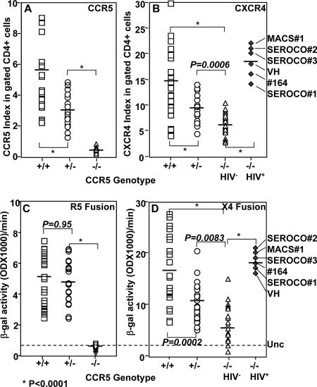
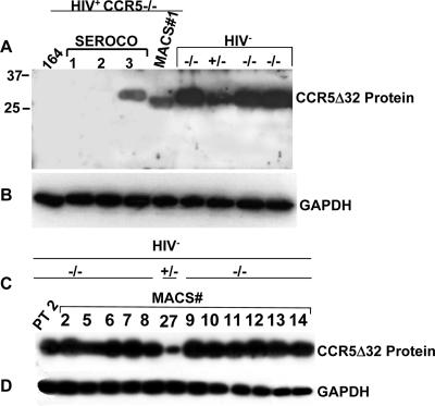
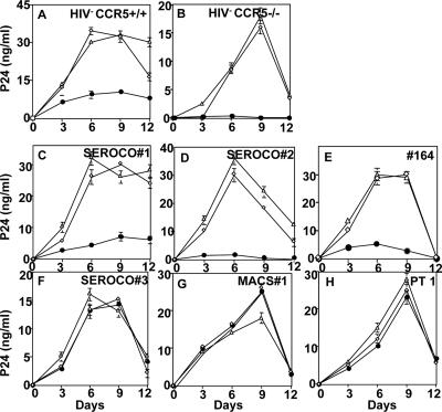
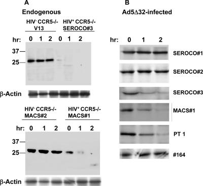
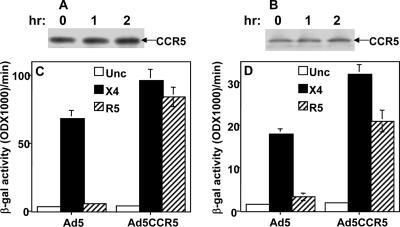
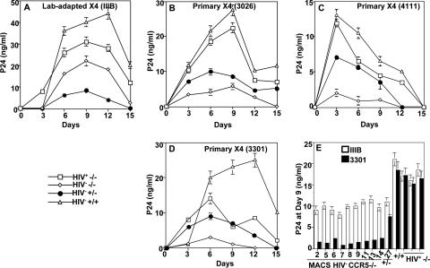
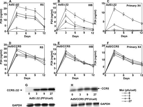
Human immunodeficiency virus type 1 (HIV-1) infection of individuals carrying the two alleles of the CCR5Delta32 mutation (CCR5(-/-)) has rarely been reported, but how the virus overcomes the CCR5Delta32 protective effect in these cases has not been delineated. We have investigated this in 6 infected (HIV(+)) and 25 HIV(-) CCR5(-/-) individuals. CD4(+) T lymphocytes isolated from HIV(-) CCR5(-/-) peripheral blood mononuclear cells (PBMCs) showed lower levels of CXCR4 expression that correlated with lower X4 Env-mediated fusion. Endogenous CCR5Delta32 protein was detected in all HIV(-) CCR5(-/-) PBMC samples (n = 25) but not in four of six unrelated HIV(+) CCR5(-/-) PBMC samples. Low levels were detected in another two HIV(+) CCR5(-/-) PBMC samples. The expression of adenovirus 5 (Ad5)-encoded CCR5Delta32 protein restored the protective effect in PBMCs from three HIV(+) CCR5(-/-) individuals but failed to restore the protective effect in PBMCs isolated from another three HIV(+) CCR5(-/-) individuals. In the latter samples, pulse-chase analyses demonstrated the disappearance of endogenous Ad5-encoded CCR5Delta32 protein and the accumulation of Ad5-encoded CCR5 during the chase periods. PBMCs isolated from CCR5(-/-) individuals showed resistance to primary X4 but were readily infected by a lab-adapted X4 strain. Low levels of Ad5-encoded CCR5Delta32 protein conferred resistance to primary X4 but not to lab-adapted X4 virus. These data provide strong support for the hypothesis that the CCR5Delta32 protein actively confers resistance to HIV-1 in vivo and suggest that the loss or reduction of CCR5Delta32 protein expression may account for HIV-1 infection of CCR5(-/-) individuals. The results also suggest that other cellular or virally induced factors may be involved in the stability of CCR5Delta32 protein.


Figures

FIG. 1.
FIG. 1.
Analysis of coreceptor expression and Env-mediated fusion of PHA-activated PBMCs isolated from CCR5+/+ (+/+), CCR5+/− (+/−), or CCR5−/− (−/−) individuals (n = 25/genotype). The PBMC samples were washed twice with PBS and then sequentially stained with PE-conjugated anti-CCR5 monoclonal antibody (3A9) or its isotype-matched control; washed and then stained with FITC-conjugated anti-CD4 monoclonal antibody or its isotype-matched control; and washed and then stained with APC-labeled anti-CXCR4 monoclonal antibody or its isotype-matched control. The MFIs of CCR5 and CXCR4 expression were measured by gating on the CD4+ population (A and B). The coreceptor expression index represents the MFI of the coreceptor staining divided by the MFI value obtained with the isotype control. (C and D) HIV-1 Env-mediated fusion with PBMC samples stained for the analysis shown in panels A and B. PHA-activated PBMCs expressing T7-lacZ were mixed with Env-expressing cells containing T7 RNA polymerase and incubated for 2.5 h at 37°C. The ability of the PBMCs to undergo cell fusion with R5 (C) or X4 (D) Env was quantified by measuring β-galactosidase (β-gal) produced. The broken line indicates the average background value obtained with the uncleaved Env (Unc) control. The indicated P values were calculated using the Student t test. The identities of the HIV+ CCR5−/− samples are indicated on the right side.
FIG. 2.
FIG. 2.
Expression analysis of CCR5Δ32 protein in infected and uninfected CCR5−/− individuals. (A) Immunodetection of native CCR5Δ32 protein in PBMCs isolated from individuals homozygous (−/−) and heterozygous (+/−) for the CCR5Δ32 mutant allele. The Western blot was probed with antibodies generated against the novel frameshift amino acids that specifically detect CCR5Δ32 protein. (B) The blot in panel A was stripped and reprobed with monoclonal antibodies to GAPDH to control for gel loading. (C) Immunodetection (same procedure as described for panel A) of native CCR5Δ32 protein in 11 CCR5−/− and 1 (+/−) PBMC samples obtained from the MACS. (D) The blot in panel C was stripped and reprobed with GAPDH antibodies. The numbers above each lane represent MACS sample numbers. PT 2, patient 2.
FIG. 3.
FIG. 3.
Rescue of the protective effect of CCR5Δ32 by Ad5-encoded CCR5Δ32 protein. The indicated PBMC samples (A to H) were infected with either Ad5 (⋄), Ad5/CCR5 (▵), or Ad5/Δ32 (•) at 50 pfu/cell for each virus, incubated for 2 days to allow protein expression, and then infected with the X4 IIIB. Culture fluids were harvested after cell suspension every 3 days and replaced with fresh medium. The amount of p24 antigen in the cell-containing supernatants was measured using an ELISA kit purchased from DuPont. The zidovudine control infection resulted in p24 values below 1 ng/ml (data not shown). Panels C to H show HIV+ CCR5−/− PBMCs. Error bars indicate standard deviations. PT 1, patient 1.
FIG. 4.
FIG. 4.
Pulse-chase analysis of CCR5Δ32 protein stability in CCR5−/− PBMCs. PHA-stimulated cells were pulse labeled with [35S]methionine and [35S]cysteine for 30 min in methionine- and cysteine-free medium DMEM containing 200 μCi of [35S]methionine and 200 μCi of [35S]cysteine per milliliter. Following the pulse, the cells were washed with FBS and reincubated in complete DMEM without radioactive label for 1 and 2 h before lysis and immunoprecipitation with rabbit antisera directed against the unique 31 amino acids of the CCR5Δ32 protein. The immunoprecipitated protein samples were analyzed on a 12.5% SDS-polyacrylamide gel. The dried gel was exposed to X-ray film and developed after 24 h. PT 1, patient 1; hr, hour.
FIG. 5.
FIG. 5.
Expression and stability of recombinant CCR5 restored R5 Env-mediated fusion with HIV+ CCR5−/− PBMC samples. (A and B) Pulse-chase analysis of CCR5 protein encoded by Ad5CCR5. The cells were coinfected with Ad5CCR5 and Ad5pT7-lacZ and used for pulse labeling. Cell lysates were prepared and immunoprecipitated with CTC-6 monoclonal antibodies to CCR5 (R&D). (C and D) HIV-1 Env-mediated fusion with two HIV+ CCR5−/− PBMC samples expressing Ad5-encoded CCR5. The PBMCs were coinfected with Ad5CCR5 and Ad5pT7-lacZ reporter. The infected cells were incubated for 48 h and then mixed with HeLa cells expressing T7 RNA polymerase and one of the indicated HIV-1 Envs. Unc is a negative Env control that has a mutation at the cleavage site and, therefore, does not promote cell fusion. The results were evaluated by measuring the β-galactosidase (β-gal) produced as a result of Env-mediated cell fusion. The PBMC samples used were MACS 1 (A and C) and SEROCO 3 (B and D). hr, hour. Error bars indicate standard deviations.
FIG. 6.
FIG. 6.
Infection kinetics of PBMCs with lab-adapted IIIB (X4) and primary X4 (isolate 4111, 3301, or 3026). Infections were performed at 10 ng/ml of the indicated X4 isolate. Culture fluid was harvested after cell resuspension every 3 days and replaced with fresh medium (A, B, C, and D). The amount of p24 antigen in the cell-containing supernatants was measured using an ELISA kit purchased from DuPont. The symbols with labeling indicate the genotype and HIV status of the PBMC sample. The MACS identity number of each PBMC sample is shown. Panel E shows the results of productive infection of the indicated PMBC samples at day 9. Error bars indicate standard deviations.
FIG. 7.
FIG. 7.
Lower expression levels of Ad5-encoded CCR5Δ32 protein induce resistance to primary X4. Affinity-purified CD4+ T lymphocytes isolated from CCR5+/+ PBMCs were used uninfected (0) or infected with increasing MOIs (3 to 27) of the indicated recombinant Ad5 viruses, incubated for 48 h to allow expression of the recombinant proteins, washed with FBS, and then infected with Ba-L (R5) (A and D), IIIB (B and E), or primary X4 (C and F). The amount of p24 antigen in the cell-containing supernatants was measured using ELISA. The zidovudine control infection resulted in p24 values below 1 ng/ml (not shown). The error bars represent replicates of the same experiment. The results shown represent one of three independent experiments using samples from three different donors. A sample of each Ad5 infection at the indicated MOI was analyzed by Western blotting to verify expression of the recombinant proteins (E and F). The CCR5 blots were performed at the same day the cells were infected with HIV-1 (day 0). The primary X4 used in these experiments was isolate 4111.

References

    1. Agrawal, L., X. Lu, Q. Jin, and G. Alkhatib. 2006. Anti-HIV therapy: current and future directions. Curr. Pharm. Des. 12:2031-2055. - PubMed
    1. Agrawal, L., X. Lu, J. Qingwen, Z. VanHorn-Ali, V. Nicolescue, D. McDermott, P. M. Murphy, and G. Alkhatib. 2004. Role for CCR5Δ32 protein in resistance to R5, R5X4, and X4 human immunodeficiency virus type 1 in primary CD4+ cells. J. Virol. 78:2277-2287. - PMC - PubMed
    1. Alkhatib, G., C. C. Broder, and E. A. Berger. 1996. Cell type-specific fusion cofactors determine human immunodeficiency virus type 1 tropism for T-cell lines versus primary macrophages. J. Virol. 70:5487-5494. - PMC - PubMed
    1. Alkhatib, G., C. Combadiere, C. C. Broder, Y. Feng, P. E. Kennedy, P. M. Murphy, and E. A. Berger. 1996. CC CKR5: a RANTES, MIP-1α, MIP-1β receptor as a fusion cofactor for macrophage-tropic HIV-1. Science 272:1955-1958. - PubMed
    1. Alkhatib, G., M. Locati, P. E. Kennedy, P. M. Murphy, and E. A. Berger. 1997. HIV-1 coreceptor activity of CCR5 and its inhibition by chemokines: independence from G protein signaling and importance of coreceptor downmodulation. Virology 234:340-348. - PubMed

Publication types