

Review
doi: 10.1128/JVI.00753-07.
Epub 2007 May 23.
Studying hepatitis C virus: making the best of a bad virus
Affiliations
- PMID: 17522203
- PMCID: PMC1951464
- DOI: 10.1128/JVI.00753-07
Item in Clipboard
Review
Studying hepatitis C virus: making the best of a bad virus
J Virol.
2007 Sep.
No abstract available
Figures
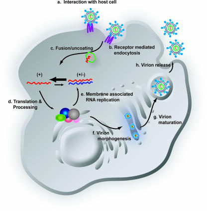
Schematic diagram of the HCV life cycle. The life cycle of HCV is similar to that of other members of the Flaviviridae family. Extracellular HCV virions interact with receptor molecules at the cell surface (a) and undergo receptor-mediated endocytosis (b) into a low-pH vesicle. Following HCV glycoprotein-mediated membrane fusion, the viral RNA is released into the cytoplasm (c). The genomic RNA is translated to generate a single large polyprotein that is processed into the 10 mature HCV proteins in association with a virus-derived ER-like membrane structure termed the membranous web (d). The mature HCV proteins replicate the RNA genome via a minus-strand replicative intermediate to produce progeny RNA. A portion of this newly synthesized RNA is packaged into nucleocapsids and associated with the HCV glycoproteins, leading to budding into the ER (f). Virions follow the cellular secretory pathway (g) and, during this transit, maturation of particles occurs (g). Mature virions are released from the cell, completing the life cycle (h). +, positive-sense genomic RNA; +/−, minus-strand replicative intermediate associated with positive-strand genomic RNA.
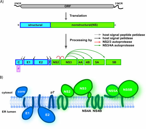
HCV genome organization, polyprotein processing, and protein topology. (A) The HCV genome is a single-stranded RNA encoding a single large open reading frame (ORF) of roughly 3,000 amino acids, flanked by structured 5′ and 3′ NCRs. The translation of the open reading frame, via the activity of an IRES element in the 5′ NCR, generates a large polyprotein that is organized with structural proteins in the amino-terminal third of the polyprotein, followed by the NS replication proteins. The polyprotein undergoes a complex co- and posttranslational series of cleavage events, catalyzed by both host and viral proteases, to produce the 10 individual HCV proteins. (B) The topology of the HCV proteins relative to the ER membrane.
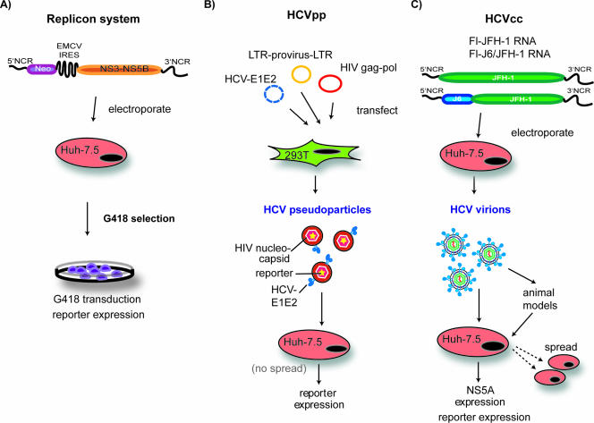
Systems for the study of HCV replication, entry, and infectivity. (A) HCV replicon systems, shown here in one of their simplest iterations, allow for productive viral RNA replication in cell culture. Bicistronic replicon RNAs, encoding a selectable marker (Neor) under control of the HCV IRES in the first cistron and the HCV replicase proteins (NS3-NS5B) under control of a heterologous IRES from encephalomyocarditis virus in the second cistron, are delivered to Huh-7-based cell lines by electroporation. Replication of these RNAs leads to production of the selectable marker and allows for selection of colonies containing active RNA replication. Transduction of resistance to the drug G418 is shown in this figure, but replicons expressing a number of reporter genes have been developed, as have methods to efficiently measure HCV proteins and RNA from these systems. (B) The HCV pseudoparticle system (HCVpp) provides a method to investigate glycoprotein-mediated events in the HCV life cycle. In this system, recombinant retroviruses that contain HCV functional glycoproteins on their surface are generated in 293T cells. These particles can be used to infect permissive cell lines, such as Huh-7.5. The retrovirus genomes have been engineered to express a reporter gene, such as luciferase, allowing for a quantitative measure of cell entry. (C) The HCVcc infectious virus system uses either JFH-1 HCV genomic RNA or chimeras of this genome with heterologous sequences (such as J6). These RNAs are electroporated into permissive cell lines and yield infectious HCV virions that can be used to infect naïve cells or animal models. Productive infection can be monitored by detection of the expression of NS5A, by a number of reporter genes, or by direct measure of viral RNA.
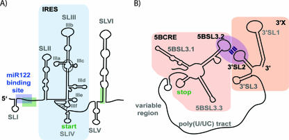
Schematic diagram of the 5′ and 3′ HCV NCRs. (A) The 5′ NCR contains six stem loop structures (SLI to SLVI). The position of the miR122 binding site within the 5′ spacer region is indicated by a blue box. The interaction site of this spacer region with the base of SLVI is also shown (green boxes). The bulk of the 5′ NCR comprises the HCV IRES element (light blue shading). The position of the four-way junction, which connects components of the SLIII loop and is involved in EIF3 binding, is indicated by a dashed circle. The pseudoknot structure within IIIf is shown by dashed lines. The position of the 5′ end of the HCV genome is shown, as is the location of the start site of polyprotein translation. (B) The 3′ NCR has a tripartite structure, containing a variable region, a poly(U/UC) tract, and the 3′ X region (further divided into 3′ stem loop structures, 3′ SL1, 3′ SL2, and 3′ SL3). The 3′ SL2 stem loop interacts via a kissing loop interaction with the 5BSL3.2 stem loop in the cis-acting RNA element in the NS5B coding sequence (5BCRE).
References
-
- Acton, S., A. Rigotti, K. T. Landschulz, S. Xu, H. H. Hobbs, and M. Krieger. 1996. Identification of scavenger receptor SR-BI as a high density lipoprotein receptor. Science 271:518-520. - PubMed
-
- Anonymous. 2002. National Institutes of Health Consensus Development Conference statement: management of hepatitis C 2002 (June 10-12, 2002). Gastroenterology 123:2082-2099. - PubMed
Publication types
MeSH terms
Substances
LinkOut - more resources
Full Text Sources
Medical
Research Materials
