. 2007 Aug;81(15):8131-9.
doi: 10.1128/JVI.00374-07. Epub 2007 May 23.

Major neutralizing sites on vaccinia virus glycoprotein B5 are exposed differently on variola virus ortholog B6

Affiliations

Major neutralizing sites on vaccinia virus glycoprotein B5 are exposed differently on variola virus ortholog B6

Lydia Aldaz-Carroll et al. J Virol. 2007 Aug.

Abstract

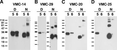
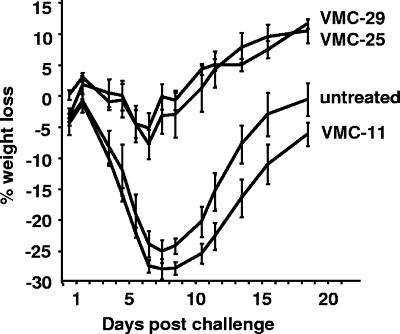
Immunization against smallpox (variola virus) with Dryvax, a live vaccinia virus (VV), was effective, but now safety is a major concern. To overcome this issue, subunit vaccines composed of VV envelope proteins from both forms of infectious virions, including the extracellular enveloped virion (EV) protein B5, are being developed. However, since B5 has 23 amino acid differences compared with its B6 variola virus homologue, B6 might be a better choice for such a strategy. Therefore, we compared the properties of both proteins using a panel of monoclonal antibodies (MAbs) to B5 that we had previously characterized and grouped according to structural and functional properties. The B6 gene was obtained from the Centers for Disease Control and Prevention, and the ectodomain was cloned and expressed in baculovirus as previously done with B5, allowing us to compare the antigenic properties of the proteins. Polyclonal antibodies to B5 or B6 cross-reacted with the heterologous protein, and 16 of 26 anti-B5 MAbs cross-reacted with B6. Importantly, 10 anti-B5 MAbs did not cross-react with B6. Of these, three have important anti-VV biologic properties, including their ability to neutralize EV infectivity and block comet formation. Here, we found that one of these three MAbs protected mice from a lethal VV challenge by passive immunization. Thus, epitopes that are present on B5 but not on B6 would generate an antibody response that would not recognize B6. Assuming that B6 contains similar variola virus-specific epitopes, our data suggest that a subunit vaccine using the variola virus homologues might exhibit improved protective efficacy against smallpox.


Figures

FIG. 1.
FIG. 1.
Production of soluble recombinant variola virus B6 in baculovirus. (A) Diagram of full-length VV B5, recombinant protein B5t generated in a previous study (1), and the recombinant variola virus B6t generated in this study. Putative transmembrane regions (TM) are shown as dashed rectangles. The signal peptide of B5 is shown as a black rectangle. Consensus N-glycosylation sites are shown as black lollipops. Numbers refer to the residues at the beginning or end of the protein or the feature depicted within the protein (e.g., TM). Additional residues appended to the recombinant protein as a result of cloning are also shown, as well as a six-histidine tag (H6). (B) Sequence alignment of B5 (WR strain; primary accession number, Q01227) and variola virus B6 (Bangladesh-1975 strain; primary accession number, Q85402). The SCRs are underlined. Residues highlighted in gray are the 23 amino acids that differ between B5 and B6, 21 of which are located in the ectodomain. Black boxes indicate putative N-glycosylation sites. The start of the transmembrane domain (TM) and cytoplasmic tail (CT) are indicated. The alignment was made using ClustalW (24).
FIG. 2.
FIG. 2.
Western blot of purified B5t and B6t. Purified baculovirus-expressed B5t (indicated by a 5) and B6t (indicated by a 6) were electrophoresed on a 12% Tris-glycine polyacrylamide gel under denaturing (D) or nonreducing (N) conditions, transferred to nitrocellulose, and probed with polyclonal IgG R182 to B5t (1 μg/ml). The film was exposed longer to show the dimer. The sizes of molecular mass markers are shown in kDa.
FIG. 3.
FIG. 3.
Inhibition of plaque formation by anti-B6 PAbs. BSC-1 cell monolayers were infected with EVs that were previously incubated for 1 h with the indicated antibody (along with anti-MV neutralizing antibodies). After 18 h of incubation at 37°C, the cells were fixed and stained with crystal violet, and plaques were counted. Data are expressed as the percentage of plaque reduction relative to the control with no IgG. Each plotted point represents an average of two wells. NRS, normal rabbit serum IgG. R195 and R196 are rabbit PAbs to B6t; PAb R182 is a rabbit PAb to B5. This experiment was done twice with the same results.
FIG. 4.
FIG. 4.
Inhibition of comet tail formation by anti-B6 PAbs. BSC-1 cell monolayers were infected with VV strain IHDJ. Following adsorption, a liquid overlay containing the indicated amount of antibody was added. After 36 h of incubation at 37°C, the cultures were fixed and stained with crystal violet and wells photographed. NRS, normal rabbit serum IgG used as a negative control; R182, a rabbit PAb against B5t used as a positive control. R195 and R196 are rabbit PAbs against B6t.
FIG. 5.
FIG. 5.
Patterns of MAb recognition of B5t and B6t by Western blotting. Purified baculovirus-expressed B5t (indicated by a 5) or B6t (indicated by a 6) was electrophoresed on a 12% Tris-glycine polyacrylamide gel under denaturing (D) or nonreducing (N) conditions, transferred to nitrocellulose and probed with each of the purified MAbs. Representative recognition patterns are shown. (A) Strong recognition of both proteins; (B) strong recognition of B6t under nonreducing conditions; (C) weak recognition of B6t under both conditions; (D) no recognition of B6t. The sizes of molecular mass markers are shown in kDa.
FIG. 6.
FIG. 6.
Percentage of blocking when each MAb is injected after initial binding of the primary antibody. X, second test antibody; Y, primary antibody. The MAbs bound to B6t with values around 200 to 300 RU, which are lower than those for the same MAbs on B5t. The MAbs are arranged according to the competition groups of B5 (1). Reciprocal blocking was done only for competing pairs. Black background, high blocking (50 to 100%); gray background, low blocking (40 to 50%); white background, no blocking. A negative value indicates increased binding of the antibody to B6t in the presence of the primary antibody.
FIG. 7.
FIG. 7.
Groupings of MAbs based on blocking interactions with B5t and B6t by biosensor. Diagrammatic representation of blocking interactions between antibodies on B5t (left) or on B6t between antibodies from the same B5t competition group (right). Blocking is represented by a black line. (A) Antibodies that react with B6t as they did on B5t; (B) antibodies that bound to B5t but behaved differently on B6t; (C) antibodies that did not bind to B5t but bound to B6t.
FIG. 8.
FIG. 8.
(A) Example of an antibody that bound to B5t but not to B6t (overlaid sensorgram)—in this case, VMC-25. (B) Example of an antibody that did not bind to B5t but bound to B6t (VMC-31).
FIG. 9.
FIG. 9.
Passive-protection studies on mice with B5t MAbs. Different anti-B5t MAbs (200 μg) were inoculated intraperitoneally into groups of five mice 24 h before intranasal challenge with approximately 1 50% lethal dose (∼5 × 10⁴ PFU) of VV. The weight of the mice was monitored, and the mean weight change ± standard error for each group is plotted. Antibodies were tested three times with similar results.

References

    1. Aldaz-Carroll, L., J. C. Whitbeck, M. Ponce de Leon, H. Lou, L. Hirao, S. N. Isaacs, B. Moss, R. J. Eisenberg, and G. H. Cohen. 2005. Epitope-mapping studies define two major neutralization sites on the vaccinia virus extracellular enveloped virus glycoprotein B5R. J. Virol. 79:6260-6271. - PMC - PubMed
    1. Aldaz-Carroll, L., J. C. Whitbeck, M. Ponce de Leon, H. Lou, L. K. Pannell, J. Lebowitz, C. Fogg, C. L. White, B. Moss, G. H. Cohen, and R. J. Eisenberg. 2005. Physical and immunological characterization of a recombinant secreted form of the membrane protein encoded by the vaccinia virus L1R gene. Virology 341:59-71. - PubMed
    1. Appleyard, G., A. J. Hapel, and E. A. Boulter. 1971. An antigenic difference between intracellular and extracellular rabbitpox virus. J. Gen. Virol. 13:9-17. - PubMed
    1. Bell, E., M. Shamim, J. C. Whitbeck, G. Sfyroera, J. D. Lambris, and S. N. Isaacs. 2004. Antibodies against the extracellular enveloped virus B5R protein are mainly responsible for the EEV neutralizing capacity of vaccinia immune globulin. Virology 325:425-431. - PubMed
    1. Boulter, E. A., and G. Appleyard. 1973. Differences between extracellular and intracellular forms of poxvirus and their implications. Prog. Med. Virol. 16:86-108. - PubMed

Publication types