. 2007 Aug;81(16):8730-41.
doi: 10.1128/JVI.00332-07. Epub 2007 May 23.

Chicken heat shock protein 90 is a component of the putative cellular receptor complex of infectious bursal disease virus

Affiliations

Chicken heat shock protein 90 is a component of the putative cellular receptor complex of infectious bursal disease virus

Ta-Wei Lin et al. J Virol. 2007 Aug.

Abstract

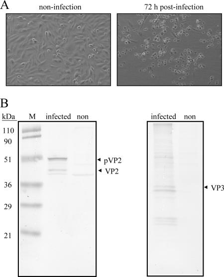
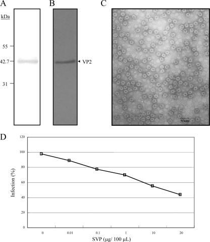
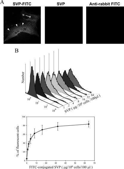
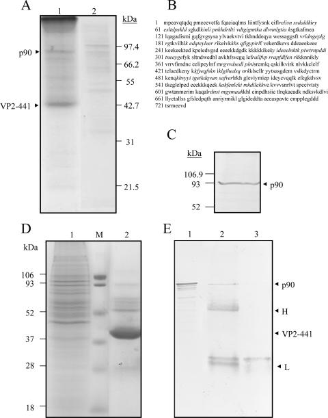
Infectious bursal disease virus (IBDV) causes a highly contagious disease in young chicks and leads to significant economic losses in the poultry industry. The capsid protein VP2 of IBDV plays an important role in virus binding and cell recognition. VP2 forms a subviral particle (SVP) with immunogenicity similar to that of the IBDV capsid. In the present study, we first showed that SVP could inhibit IBDV infection to an IBDV-susceptible cell line, DF-1 cells, in a dose-dependent manner. Second, the localizations of the SVP on the surface of DF-1 cells were confirmed by fluorescence microscopy, and the specific binding of the SVP to DF-1 cells occurred in a dose-dependent manner. Furthermore, the attachment of SVP to DF-1 cells was inhibited by an SVP-induced neutralizing monoclonal antibody against IBDV but not by denatured-VP2-induced polyclonal antibodies. Third, the cellular factors in DF-1 cells involved in the attachment of SVP were purified by affinity chromatography using SVP bound on the immobilized Ni(2+) ions. A dominant factor was identified as being chicken heat shock protein 90 (Hsp90) (cHsp90) by mass spectrometry. Results of biotinylation experiments and indirect fluorescence assays indicated that cHsp90 is located on the surface of DF-1 cells. Virus overlay protein binding assays and far-Western assays also concluded that cHsp90 interacts with IBDV and SVP, respectively. Finally, both Hsp90 and anti-Hsp90 can inhibit the infection of DF-1 cells by IBDV. Taken together, for the first time, our results suggest that cHsp90 is part of the putative cellular receptor complex essential for IBDV entry into DF-1 cells.


Figures

FIG. 1.
FIG. 1.
Infection of DF-1 cells with a local IBDV strain, strain P3009. (A) CPE of DF-1 cells caused by IBDV infection. More cells with a round shape (CPE) (right) were seen, and some infected cells died 72 h postinfection; cells in the noninfected group were still healthy (left). (B) Western blot of IBDV-infected DF-1 cells. Two major structural proteins, pVP2/VP2 and VP3, with molecular masses of 50/43 kDa and 33 kDa, respectively, generated in the IBDV-infected DF-1 cells were recognized by anti-VP2 (left) and anti-VP3 (right) polyclonal antibodies, respectively, and neither pVP2/VP2 nor VP3 was observed in the noninfected group. Molecular mass standards are shown on the left in kDa.
FIG. 2.
FIG. 2.
Purity analysis of VP2-441 SVP prepared by IMAC and inhibition of IBDV infection of DF-1 cells by SVP. IMAC-purified SVP was concentrated through a centrifugal filter (100 kDa) and analyzed by SDS-PAGE with silver staining (A) and Western blotting with anti-VP2 polyclonal antibody (B). Molecular mass standards are shown on the left in kDa. (C) Electron micrograph with ×100,000 magnification (scale bar, 50 nm) and negatively stained with 2% uranyl acetate. (D) Dose-dependent inhibition of IBDV infection by SVP. DF-1 cells were preincubated with different concentrations of SVP (0, 0.01, 0.1, 1, 10, and 20 μg/100 μl) for 1 h at 4°C and then infected with 10 TCID50 of IBDV P3009 for 1 h. CPE was determined using crystal violet staining 96 h after infection. Two separate experiments showed similar results.
FIG. 3.
FIG. 3.
Characterization of SVP binding to DF-1 cells by confocal microscopy (A) and flow cytometry (B). DF-1 cells grown on microscope slides were incubated with FITC-labeled SVP (left), unlabeled SVP (middle), and FITC-labeled goat anti-rabbit IgG (right) for 60 min at 4°C. Cells were fixed in 4% paraformaldehyde for 60 min after the removal of unbound SVP and antibody. Cells were examined using a confocal microscope (Zeiss LSM510) at ×630 magnification or a fluorescent microscope (Nikon TS-2000) at ×400 magnification. The cell nucleus (N) and the represented endosome (arrows) are indicated. (B) Saturation of SVP binding to DF-1 cells. Aliquots of 106 DF-1 cells incubated with increasing amounts of the FITC-conjugated SVPs were submitted to one-color fluorescence flow cytometric analysis. A typical flow cytometry histogram is shown on the top. The percentage of cell-bound SVP (FITC-positive cells) was determined in three separate experiments. Average values are shown with standard deviations (bottom).
FIG. 4.
FIG. 4.
Dose-dependent inhibition of SVP binding to DF-1 cells by MAbSVP-4, a neutralizing MAb. (A) MAbSVP-4 protects CEF cells from IBDV infection. The IBDV neutralization capabilities of MAbSVP-4, MAbSVP-1, and anti-VP2 were measured by virus neutralization assays. Serum samples were diluted twofold, starting at a 1:16 dilution, in Medium 199 and mixed with 200 TCID50 of IBDV P3009 per well (final serum dilution, 1:10,240) at 37°C for 1 h. The antibody-virus mixture was then added to 2 × 10⁴ CEF cells in Medium 199 containing 10% fetal calf serum per well in a 96-well culture plate. After 4 days at 37°C, cell monolayers were observed under a microscope for CPE and were then washed with PBS and stained for 20 min with 1.5% crystal violet in 50% ethanol. The titer of neutralizing activity was evaluated by visual screening of the infected monolayers, and the end point was calculated as the reciprocal value of the highest serum dilution that causes a 50% reduction of the cell monolayer. Results for MAbSVP-4 with a dilution factor of 1,024 and for MAbSVP-1 with a dilution factor of 16 (same as anti-VP2) are shown. CPE of cells incubated with a mixture of MAbSVP-1 (or anti-VP2) and virus could be observed as clearly as that in the negative control, where CEF cells were infected with only 200 TCID50 of the virus. A similar result was not shown for the cells treated with a mixture of anti-VP2 and virus. In contrast, with MAbSVP-4 neutralizing IBDV and protecting the cells from the infection, cells incubated with MAbSVP-4 and virus complex were as healthy as cells of the control group (top). (B) Flow cytometry histograms. Shown are staining DF-1 cells with FITC-conjugated SVP preincubated with either different concentrations of MAbSVP-4 or anti-VP2. Violet, control cells; blue, cells incubated with FITC-labeled SVP; red, cells incubated with FITC-labeled SVP and MAbSVP-4; green, cells incubated with FITC-labeled SVP and anti-VP2 polyclonal antibody.
FIG. 5.
FIG. 5.
Isolation of cHsp90 with affinity for SVP. (A) Affinity isolation of a p90 protein with SVP as the IMAC ligand. A procedure described in the text was set up for affinity isolation of putative IBDV receptors using SVP as the ligand bound to the immobilized Ni2+ ions. In brief, total proteins from DF-1 cells were passed through the affinity chromatographic column. After washing with two wash buffers, the elution of SVP and its associated factors was accomplished by using an IMAC elution buffer. The fractions collected were concentrated, and 40 μl of the concentrate (lane1) and, as a control, 50 μl of the concentrated elution fraction obtained in the absence of SVP (lane 2) were separated by SDS-12.5% PAGE and stained with Coomassie blue (left). Positions of a p90 protein and the monomer VP2-441 that forms SVP are indicated on the left. (B) Identification of p90 by mass spectrometry. The protein sequence of cHsp90 was derived from Protein Data Bank accession number P11501. Peptide sequences of p90 identified by mass spectrometry are shown in italics. (C) Identification of p90 as cHsp90 by Western blotting. An aliquot of 40 μl of the concentrated elution from the affinity column chromatography with SVP was separated by SDS-12.5% PAGE, transferred onto a PVDF membrane, and incubated with an anti-Hsp90 MAb. The membranes were then incubated with a second antibody coupled to alkaline phosphatase and developed in a buffer containing NBT and BCIP. Molecular mass markers are indicated on the left. An arrow indicates the position of p90 (cHsp90). (D) Isolation of p90 from DF-1 cells by immunoprecipitation. Total proteins from DF-1 cells (lane 1) and the precipitated immune complexes (lane 2) were separated by SDS-12.5% PAGE and stained with Coomassie blue (left). The precipitated immune complexes in the presence of SVP (lane 2) and without SVP (lane 3) were separated by SDS-12.5% PAGE, transferred onto a PVDF membrane, and incubated with anti-Hsp90. (E) Western blots were developed as described above. As expected, cHsp90 (p90) was identified in the total DF-1 lysate (lane 1). Molecular mass markers are indicated on the left. Arrows indicate positions of proteins of the immune complexes.
FIG. 6.
FIG. 6.
Surface localization of the 90-kDa protein from DF-1 cells. (A) Purification of biotinylated membrane-bound cHsp90 of DF-1 cells by SVP. Surface-biotinylated proteins from DF-1 were passed through a column with SVPs coupled to Ni-NTA-Sepharose. Aliquots of 40 μl of the elution fraction (lane 1), the elution fractions obtained in the absence of SVP (as controls) (lane 2), and a biotinylated recombinant Hsp90 (lane Biotin-Hsp90) were separated by SDS-12.5% PAGE, transferred onto a PVDF membrane, and stained with anti-Hsp90 MAb and a proper secondary antibody (left) or streptavidin-alkaline phosphatase (AP) (right), NBT, and BCIP. Molecular mass markers are indicated on the left. The arrow indicates the migration of eluted p90 present in DF-1 cells. An unidentified product has also been visualized. (B) Localization of cHsp90 on the surface of DF-1 by indirect immunofluorescence. Nonpermeabilized cells were incubated with anti-β-actin MAb (middle) or anti-Hsp90 (right), followed by staining with a proper secondary antibody, and examined by fluorescence microscopy at ×600 magnification. A phase-contrast image of cells is shown on the left. (C) Characteristic distribution of cHsp90 among the PM of DF-1 cells. DF-1 cells grown on microscope slides were prefixed and then incubated with FITC-labeled SVP or treated with anti-Hsp90 as described above. After mounting, cells were examined using a confocal microscope (FluoView FV1000; Olympus). Characteristic distributions of SVP (top) and cHsp90 (bottom) among the PM are indicated by arrows. Differential interference contrast (DIC) was used to yield a better image when viewed in bright-field illumination. Bar, 10 μm.
FIG. 7.
FIG. 7.
VOPBA and far-Western assay of p90 and infection inhibition assays of recombinant Hsp90 protein and anti-Hsp90 in DF-1 cells. Elution fractions containing proteins from DF-1 showing affinity to SVP bound to the immobilized Ni2+ ions were separated by SDS-12.5% PAGE and transferred onto PVDF membranes. Membranes were incubated with (A) or without (C) 2 × 10⁴ PFU of IBDV and later with a rabbit polyclonal anti-VP2 antibody and goat anti-rabbit IgG coupled to alkaline phosphatase; color was developed with BCIP and NBT. A similar procedure was performed for the far-Western assay (B), except that IBDV was replaced with SVP. Molecular mass markers are indicated on the right. p90 (marked with an arrow) from DF-1 cells was recognized by IBDV (A) and SVP (B). Notably, some other proteins were slightly recognized. (D) Infection inhibition assays with anti-Hsp90 antibody. DF-1 cells were preincubated with different concentrations of SVPMAb-1 (square, mouse monoclonal antibody as a control) or anti-Hsp90 antibody (triangle) for 1 h at 4°C. Subsequently, cells were infected with 100 TCID50 of IBDV. Culture supernatants were collected 96 h after infection, and the resulting infectious virus titer was determined. Each point represents the average of two separate experiments. (E) Infection inhibition assays of recombinant Hsp90. Recombinant Hsp90 protein (Hsp90α) of different concentrations or BSA (as a negative control) was preincubated with 100 TCID50 of IBDV for 1 h at 39°C prior to its incubation with DF-1 cells. Culture supernatants were collected 96 h after infection, and the resulting infectious virus titer was determined. Each point represents the average of two separate experiments.

Similar articles

Cited by

References

    1. Bass, D. M., and H. B. Greenberg. 1992. Strategies for the identification of icosahedral virus receptors. J. Clin. Investig. 89:3-9. - PMC - PubMed
    1. Bavari, S., C. M. Bosio, E. Wiegand, G. Ruthel, A. B. Will, T. W. Geisbert, M. Hevey, C. Schmaljohn, A. Schmaljohn, and M. J. Aman. 2002. Lipid raft microdomains: a gateway for compartmentalized trafficking of Ebola and Marburg viruses. J. Exp. Med. 195:593-602. - PMC - PubMed
    1. Byrd, C. A., W. Bornmann, H. Erdjument-Bromage, P. Tempst, N. Pavletich, N. Rosen, C. F. Nathan, and A. Ding. 1999. Heat shock protein 90 mediates macrophage activation by Taxol and bacterial lipopolysaccharide. Proc. Natl. Acad. Sci. USA 96:5645-5650. - PMC - PubMed
    1. Chen, C. S., S. Y. Suen, S. Y. Lai, G. R. Chang, T. C. Lu, M. S. Lee, and M. Y. Wang. 2005. Purification of capsid-like particles of infectious bursal disease virus (IBDV) VP2 expressed in E. coli with a metal-ion affinity membrane system. J. Virol. Methods 130:51-58. - PubMed
    1. Cheng, Y. S., M. S. Lee, S. Y. Lai, S. R. Doong, and M. Y. Wang. 2001. Separation of pure and immunoreactive virus-like particles using gel filtration chromatography following immobilized metal ion affinity chromatography. Biotechnol. Prog. 17:318-325. - PubMed

Publication types

MeSH terms

Substances

LinkOut - more resources