. 2007 Aug;81(15):7873-84.
doi: 10.1128/JVI.01845-06. Epub 2007 May 23.

Human immunodeficiency virus type 1 assembly, budding, and cell-cell spread in T cells take place in tetraspanin-enriched plasma membrane domains

Affiliations

Human immunodeficiency virus type 1 assembly, budding, and cell-cell spread in T cells take place in tetraspanin-enriched plasma membrane domains

Clare Jolly et al. J Virol. 2007 Aug.

Abstract

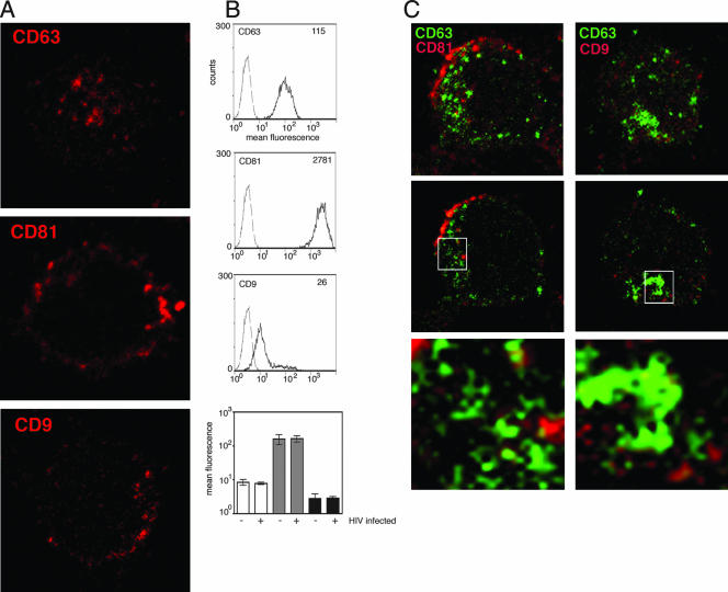
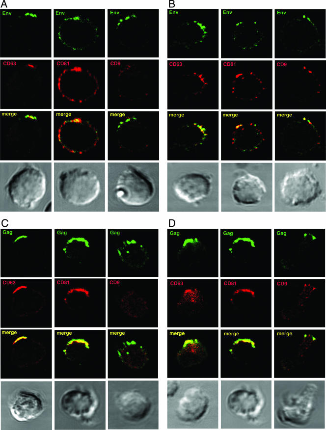
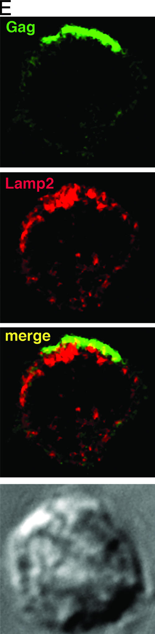
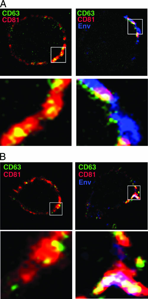
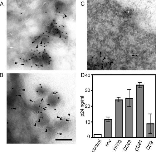
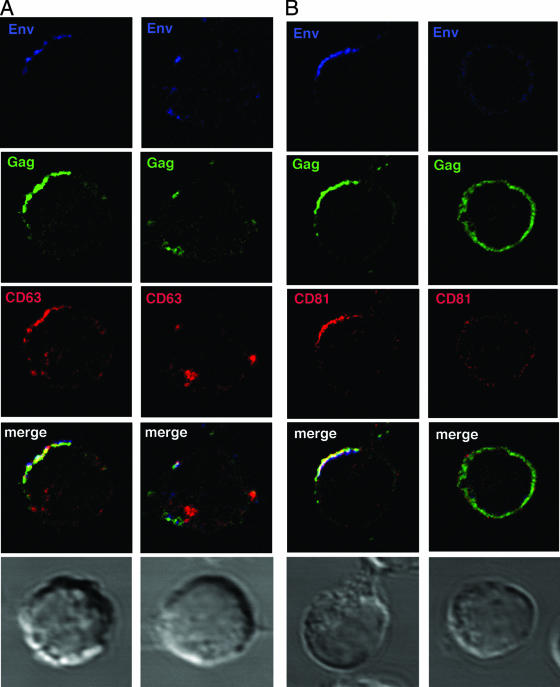
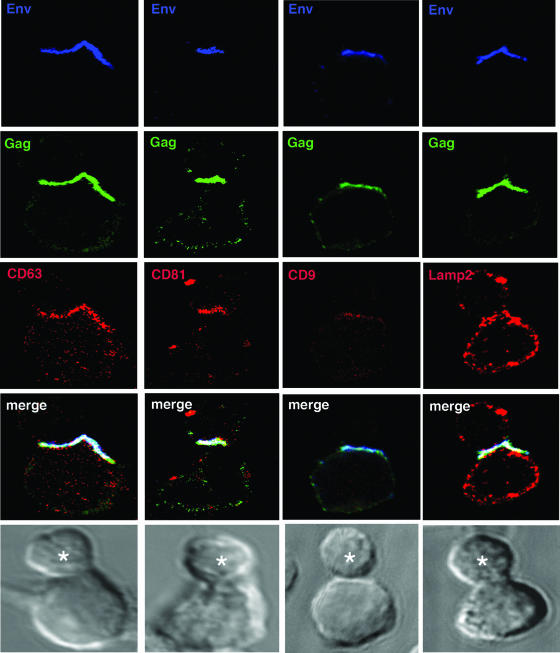
Human immunodeficiency virus type-1 (HIV-1) egress from infected CD4+ T cells is thought to be via assembly and budding at the plasma membrane and may involve components of the T-cell secretory apparatus, including tetraspanins. However, many studies on HIV-1 assembly have examined the trafficking of viral proteins in isolation, and most have used immortalized epithelial, fibroblastic, or hematopoietic cell lines that may not necessarily reflect natural infection of susceptible T cells. Here we have used immunofluorescence and cryoimmunoelectron microscopy (CEM) to examine protein transport during HIV-1 assembly in productively infected Jurkat CD4+ T cells and primary CD4+ T cells. The HIV-1 envelope glycoprotein (Env) and the core protein (Gag) colocalize strongly with CD63 and CD81 and less strongly with CD9, whereas no colocalization was seen between Env or Gag and the late endosome/lysosomal marker Lamp2. CEM revealed incorporation of CD63 and CD81 but not Lamp2 into virions budding at the plasma membrane, and this was supported by immunoprecipitation studies, confirming that HIV-1 egress in T cells is trafficked via tetraspanin-enriched membrane domains (TEMs) that are distinct from lysosomal compartments. CD63, CD81, and, to a lesser extent, CD9 were recruited to the virological synapse (VS), and antibodies against these tetraspanins reduced VS formation. We propose that HIV-1 promotes virus assembly and cell-cell transfer in T cells by targeting plasma membrane TEMs.


Figures

FIG. 1.
FIG. 1.
Distribution of tetraspanins in uninfected Jurkat T cells. (A) Uninfected Jurkat T cells were adhered to poly-l-lysine-coated coverslips for 1 h at 37°C, fixed with paraformaldehyde, permeabilized, and stained with MAbs against CD63, CD81, or CD9 (red). Images shown are single confocal sections through the middle of a cell. (B) Cell surface expression of CD63, CD81, and CD9 was measured on uninfected Jurkat T cells by flow cytometry. The mean fluorescence intensity of CD63, CD81, and CD9 staining (solid line) is shown compared to a control MAb (broken line), and the mean fluorescence values (insert) are shown for each plot. Levels of surface expression of CD63 (white bars), CD81 (gray bars), and CD9 (black bars) are compared between HIV-1 infected Jurkat cells and uninfected Jurkat cells (bottom panel). Data are the means of three independent experiments, and error bars represent the standard error of the mean. (C) CD63 does not colocalize with CD81 or CD9 in Jurkat T cells. Jurkat cells were adhered to coverslips, fixed, permeabilized, and stained for CD63 (green) and either CD81 (left panel, red) or CD9 (right panel, red). Primary MAbs were detected with anti-IgG2b (CD63) and anti-IgG1 (CD81 and CD9) isotype-specific conjugated secondary antibodies. The three-dimensional reconstructed z series (top) and a single optical section through the middle of the cell (center) are shown. The bottom panels each show a magnified image of a region from the single optical section selected for strong staining (boxed).
FIG. 2.
FIG. 2.
HIV-1 Env and Gag colocalize with tetraspanins in virus-infected T cells. HIV-1-infected JurkatLAI T cells (A) or primaryLAI cells (B) were surface stained for Env and tetraspanins. Cells were adhered to coverslips for 1 h at 37°C in the presence of the human Env-specific antibody 2G12 and then incubated at 4°C for 30 min with MAbs against CD63 (left panel), CD81 (center panel), or CD9 (right panel). Cells were fixed, and surface staining of Env (green) and tetraspanins (red) was analyzed by LSCM. Images are single sections through the middle of a cell, and areas of colocalization are yellow. JurkatLAI T cells (C) or primaryLAI cells (D) were permeabilized and stained for Gag and tetraspanins. Cells were adhered to coverslips, fixed and permeabilized, and incubated with rabbit antisera against Gag p17 and p24 and MAbs against CD63 (left panel), CD81 (center panel), or CD9 (right panel). Intracellular staining of Gag (green) and tetraspanins (red) was analyzed by LSCM. Images are single sections through the middle of the cell, and areas of colocalization are yellow. (E) Jurkat LAI cells were adhered to coverslips, fixed, permeabilized, and stained for intracellular Gag and Lamp2. Images are single sections through the middle of the cell, and areas of colocalization are yellow.
FIG. 2.
FIG. 2.
HIV-1 Env and Gag colocalize with tetraspanins in virus-infected T cells. HIV-1-infected JurkatLAI T cells (A) or primaryLAI cells (B) were surface stained for Env and tetraspanins. Cells were adhered to coverslips for 1 h at 37°C in the presence of the human Env-specific antibody 2G12 and then incubated at 4°C for 30 min with MAbs against CD63 (left panel), CD81 (center panel), or CD9 (right panel). Cells were fixed, and surface staining of Env (green) and tetraspanins (red) was analyzed by LSCM. Images are single sections through the middle of a cell, and areas of colocalization are yellow. JurkatLAI T cells (C) or primaryLAI cells (D) were permeabilized and stained for Gag and tetraspanins. Cells were adhered to coverslips, fixed and permeabilized, and incubated with rabbit antisera against Gag p17 and p24 and MAbs against CD63 (left panel), CD81 (center panel), or CD9 (right panel). Intracellular staining of Gag (green) and tetraspanins (red) was analyzed by LSCM. Images are single sections through the middle of the cell, and areas of colocalization are yellow. (E) Jurkat LAI cells were adhered to coverslips, fixed, permeabilized, and stained for intracellular Gag and Lamp2. Images are single sections through the middle of the cell, and areas of colocalization are yellow.
FIG. 3.
FIG. 3.
HIV-1 Env accumulates in CD63+ CD81+ TEMs at the plasma membrane of T cells. (A) Jurkat T cells and (B) primary CD4+ T cells were used uninfected (left panel) or infected with HIV-1 (right panel). Cells were adhered to coverslips for 1 h at 37°C in the presence of the human Env-specific antibody 2G12 and then incubated at 4°C for 30 min with MAbs against CD63 and CD81. Cells were fixed, and the surface staining of Env (blue), CD63 (green), and CD81 (red) was analyzed by LSCM. The top panels show a single section through the middle of the cell, and the bottom panels are magnified images of a region selected for strong staining from the single optical section (boxed). Regions of CD63 and CD81 colocalization are yellow, and regions of Env, CD63, and CD81 triple colocalization appear white.
FIG. 4.
FIG. 4.
CD63 and CD81 are incorporated into budding virions. Ultrathin cryosections of HIV-1-infected JurkatLAI cells were labeled with rabbit antiserum against HIV Gag p17 and a mouse MAb specific for either CD63 (A) or CD81 (B). Primary antibodies were labeled with mouse-specific 5-nm and rabbit-specific 10-nm gold colloids. CD63 and CD81 labeling (5-nm gold colloid) is highlighted with arrowheads. (C) Cryosections were labeled with a mouse MAb against Gag and rabbit antiserum specific for Lamp2. Here Gag was visualized with mouse-specific 5-nm gold colloids and Lamp2 with rabbit-specific 10-nm colloids. Lamp2 labeling is highlighted with arrowheads, and a budding virion (Lamp Gag+) is highlighted with an asterisk. Bar, 100 nm. (D) Virus precipitation with antibodies against HIV-1 proteins or tetraspanins. Cell-free viral supernatants were precipitated with Env-specific antibodies (MAbs 2G12 and IgG1b12), tetraspanin-specific MAbs, or a control antibody, and the Gag p24 content of the precipitated material was measured by ELISA. The nonspecific background signal was determined by incubating viral supernatants with preblocked Pansorbin cells without antibody, and these values were subtracted to calculate the concentration of p24 in ng/ml. Bars represent means of two independent experiments, and variability is represented by the standard error of the mean.
FIG. 5.
FIG. 5.
Actin depolymerization disrupts TEM-HIV-1 polarized assembly platforms. JurkatLAI cells (5 × 10⁵) were washed, resuspended in RPMI 1640-1% FCS, and adhered to poly-l-lysine-coated coverslips at 37°C. The cells were either untreated (left panel) or treated with latrunculin A (right panels) and stained for HIV-1 Env with the MAb 50-69 (blue). Cells were fixed, permeabilized, and stained for HIV-1 Gag (green) and either CD63 (A [red]) or CD81 (B [red]). Images are single sections through the middle of the cell with the corresponding Nomarski image: areas of red/green colocalization appear yellow, and areas of red/green/blue colocalization appear white.
FIG. 6.
FIG. 6.
Tetraspanins are components of the T-cell VS. HIV-1-infected JurkatLAI cells (effectors) were mixed with an equal number of freshly-isolated primary CD4+ T cells (targets) and incubated on poly-l-lysine-treated coverslips in the presence of the Env-specific MAb 50-69 (blue) for 1 h at 37°C. Conjugate evolution was arrested by fixing, and cells were permeabilized and stained using rabbit antisera directed against Gag p17 and p24 (green) and mouse MAbs specific for either CD63 (red, left panel), CD81 (red, second panel from left), or CD9 (red, second panel from right). Alternatively, cells were stained with a mouse MAb against Gag (green) and rabbit antisera against Lamp2 (red, right panel). CD81 and CD63, and to a lesser extent CD9, but not Lamp2, are enriched in the effector cell at the conjugate interface and colocalize with HIV-1 Env and Gag. Images are single sections through the middle of a conjugate, and the target cell is labeled with an asterisk. Areas of colocalization appear white, and the corresponding Nomarski image is shown.

References

    1. Batonick, M., M. Favre, M. Boge, P. Spearman, S. Honing, and M. Thali. 2005. Interaction of HIV-1 Gag with the clathrin-associated adaptor AP-2. Virology 342:190-200. - PubMed
    1. Berditchevski, F., and E. Odintsova. 1999. Characterization of integrin-tetraspanin adhesion complexes: role of tetraspanins in integrin signaling. J. Cell Biol. 146:477-492. - PMC - PubMed
    1. Bhattacharya, J., P. J. Peters, and P. R. Clapham. 2004. Human immunodeficiency virus type 1 envelope glycoproteins that lack cytoplasmic domain cysteines: impact on association with membrane lipid rafts and incorporation onto budding virus particles. J. Virol. 78:5500-5506. - PMC - PubMed
    1. Boge, M., S. Wyss, J. S. Bonifacino, and M. Thali. 1998. A membrane-proximal tyrosine-based signal mediates internalization of the HIV-1 envelope glycoprotein via interaction with the AP-2 clathrin adaptor. J. Biol. Chem. 273:15773-15778. - PubMed
    1. Booth, A. M., Y. Fang, J. K. Fallon, J. M. Yang, J. E. Hildreth, and S. J. Gould. 2006. Exosomes and HIV Gag bud from endosome-like domains of the T cell plasma membrane. J. Cell Biol. 172:923-935. - PMC - PubMed

Publication types

MeSH terms