. 2007 Aug;81(15):8236-46.
doi: 10.1128/JVI.02694-06. Epub 2007 May 23.

Human immunodeficiency virus type 1 Vif inhibits packaging and antiviral activity of a degradation-resistant APOBEC3G variant

Affiliations

Human immunodeficiency virus type 1 Vif inhibits packaging and antiviral activity of a degradation-resistant APOBEC3G variant

Sandrine Opi et al. J Virol. 2007 Aug.

Abstract

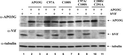
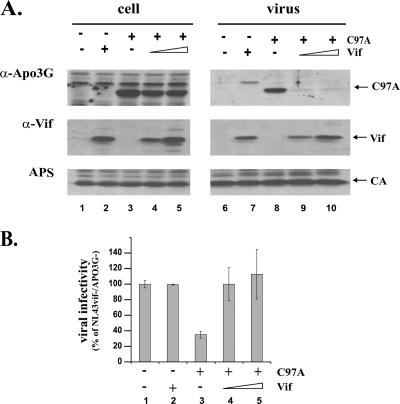
Human immunodeficiency virus type 1 (HIV-1) Vif counteracts the antiviral activity of the human cytidine deaminase APOBEC3G (APO3G) by inhibiting its incorporation into virions. This has been attributed to the Vif-induced degradation of APO3G by cytoplasmic proteasomes. We recently demonstrated that although APO3G has a natural tendency to form RNA-dependent homo-multimers, multimerization was not essential for encapsidation into HIV-1 virions or antiviral activity. We now demonstrate that a multimerization-defective APO3G variant (APO3G C97A) is able to assemble into RNase-sensitive high-molecular-mass (HMM) complexes, suggesting that homo-multimerization of APO3G and assembly into HMM complexes are unrelated RNA-dependent processes. Interestingly, APO3G C97A was highly resistant to Vif-induced degradation even though the two proteins were found to interact in coimmunoprecipitation experiments and exhibited partial colocalization in transfected HeLa cells. Surprisingly, encapsidation and antiviral activity of APO3G C97A were both inhibited by Vif despite resistance to degradation. These results demonstrate that targeting of APO3G to proteasome degradation and interference with viral encapsidation are distinct functional properties of Vif.


Figures

FIG. 1.
FIG. 1.
APO3G C97A is resistant to Vif-induced degradation. (A) HeLa cells were transfected with vectors expressing untagged wt APO3G (lanes 2 to 4) or APO3G C97A (lanes 5 to 7) together with vif-deficient pNL-A1 (lanes 2 and 5) or increasing amounts of the Vif-expressing pNL-A1 vector as described in the text (lanes 3, 4, 6, and 7). Lane 1 is a control that expresses Vif in the absence of APO3G. The total amount of transfected DNA in each sample was adjusted to 5 μg using empty pcDNA3.1 vector DNA (lane 1) or pNL-A1vif(−) DNA (lanes 2 to 7). Cells were harvested 24 h after transfection, and whole-cell lysates were analyzed by immunoblotting using an APO3G-specific rabbit polyclonal antibody (ApoC17) followed by incubation with an HRP-conjugated anti-rabbit antibody (APO3G). The same blot was subsequently reblotted with a Vif-specific monoclonal antibody (Vif), followed by probing with an antibody to α-tubulin (tubulin). Proteins are identified on the right. APO3G-specific protein bands were quantified by densitometric scanning of the gel, and signal intensities were calculated as percentages of the signal observed in the absence of Vif (right side). (B) HeLa cells were transfected and analyzed as for panel A, except that pNL-A1 was replaced by the pcDNA-hVif vector carrying a codon-optimized vif gene. DNA amounts were adjusted to 5 μg using empty pcDNA3.1 vector DNA. (C) HeLa cells (5 × 10⁶) were transfected with constant amounts of myc-tagged wt pcDNA-APO3Gmyc and untagged pcDNA-APO3G C97A together with increasing amounts of pcDNA-hVif. Plasmid ratios of hVif/APO3G vectors were 0 (lane 2), 0.25 (lane 3), and 0.5 (lane 4). Total amounts of DNA were adjusted to 5 μg by addition of empty-vector DNA (pcDNA3.1) as appropriate. A mock-transfected sample (no DNA) was included as a control (lane 1). Whole-cell lysates were prepared 24 h after transfection and subjected to immunoblotting using an APO3G-specific peptide antibody (top) or a Vif monoclonal antibody (bottom). Proteins are identified on the right. Quantitation of APO3G-specific bands for panels B and C was done as for panel A.
FIG. 2.
FIG. 2.
APO3G C97A binds to Vif. (A) HeLa cells were cotransfected with pcDNA-hVif and myc-tagged wt pcDNA-APO3Gmyc (lanes 2 and 5) or myc-tagged pcDNA-APO3G C97A-myc (lane 3 and 6). To prevent degradation of wt APO3G by Vif, the dominant-negative Cul5 mutant pCul5-RbxHA was cotransfected in all samples. Total DNA amounts were adjusted to 5 μg in each sample by using pcDNA3.1 vector DNA. As a control, HeLa cells were transfected with pcDNA-hVif and pCul5-RbxHA in the absence of APO3G (lanes 1 and 3). Samples were analyzed either directly (left panels) or following immunoprecipitation (IP) with a myc-specific polyclonal antibody (right panels). Cell lysates and immunoprecipitates were analyzed by immunoblotting using an APO3G-specific rabbit polyclonal antibody, followed by incubation with an HRP-conjugated anti-rabbit antibody (top panels), or a monoclonal antibody to Vif, followed by incubation with an HRP-conjugated anti-mouse antibody (bottom panels). Proteins are identified on the right. In the immunoprecipitated samples, the HRP-conjugated second antibody reacted with both the APO3G-specific antibodies and the myc-specific antibody used for the immunoprecipitation (immunoglobulin G [IgG]). (B) The relative binding efficiencies of Vif and APO3G were determined by quantifying the efficiency of Vif coimmunoprecipitation with APO3G. APO3G- and Vif-specific protein bands in the right panels of panel A (IP) were quantified by densitometric scanning. Signals were corrected for differences in the amounts of immunoprecipitated APO3G protein. The signal intensity of Vif coprecipitated with wt APO3G was defined as 100%. Error bars in the APO3G C97A sample (C97A) reflect the standard deviation calculated from three independent co-IP studies.
FIG. 3.
FIG. 3.
APO3G C97A is resistant to Vif from HIV-1, SIVagm, and SIVmac. HeLa cells were transfected with vectors expressing wt APO3G (C and D) or APO3G C97A (A and B) together with vif-deficient pNL-A1 (lane 1), pNL-A1 (lane 2), pNL-A1/AgmVif (lane 3), or pNL-A1/MacVif (lane 4). Whole-cell lysates were analyzed for the expression of APO3G by using a rabbit polyclonal peptide antibody (B and D) or a mixture of antibodies to Vif consisting of a monoclonal antibody to HIV-1 Vif plus polyclonal rabbit antibodies to SIVagm Vif and SIVmac Vif (A and C). Vif-specific antibodies were subsequently identified with a mixture of HRP-conjugated anti-mouse and anti-rabbit antibodies. Proteins are identified on the right.
FIG. 4.
FIG. 4.
Mutation of cysteine residues in the N-terminal but not the C-terminal zinc finger motif of APO3G confers resistance to Vif. HeLa cells were transfected with pcDNA-APO3G (lanes 2 and 3), pcDNA-APO3G C97A (lanes 4 and 5), pcDNA-APO3G C100S (lanes 6 and 7), pcDNA-APO3G C97A/C100S (lanes 8 and 9), or pcDNA-APO3G C288S/C291A (lanes 10 and 11) in the absence (even-numbered lanes) or presence (odd-numbered lanes) of pcDNA-hVif plasmid DNA at a 1:1 ratio. All APO3G vectors encoded untagged APO3G proteins. A sample transfected with pcDNA-hVif in the absence of APO3G was included as a control (lane 1). Total DNA amounts were adjusted to 5 μg in each sample by using empty pcDNA3.1 vector DNA. Cells were harvested 24 h after transfection and subjected to immunoblotting as described for Fig. 2.
FIG. 5.
FIG. 5.
Intracellular distribution of APO3G C97A. (A to D) HeLa cells were transfected with equal amounts of APOBEC3G-HA and pcDNA-APO3G C97Amyc vector DNA. Transfected cells were grown in 12-well plates on coverslips. Cells were fixed with methanol 20 h after being transfected and reacted with an HA-specific mouse monoclonal antibody and a myc-specific polyclonal rabbit antibody, followed by incubation with Cy2-conjugated donkey anti-mouse and Texas Red-conjugated donkey anti-rabbit antibodies. All antibodies were used at 1:200 dilutions. Images of Cy2-stained wt APO3G (A) and Texas Red-stained APO3G C97A (B) were acquired simultaneously and stored in separate image channels. Bright-field (Nomarski) images were stored in a third image channel (C). Colocalization of wt and mutant APO3G was determined by combining the green and red image channels (D). (E to H) HeLa cells were transfected with untagged pcDNA-APO3G C97A and pNL-A1 plasmid DNAs at 1:0.5 molar ratios. Cells were processed for image analysis as described for panels A to D. Cells were stained with a Vif-specific mouse monoclonal antibody and an APO3G-specific rabbit polyclonal antibody. Cy2 and Texas Red-conjugated secondary antibodies were as for panels A to D. Images of Cy2-stained Vif (E) and Texas Red-stained APO3G C97A (F) were acquired simultaneously and stored in separate image channels. Bright-field (Nomarski) images were stored in a third image channel (G). Colocalization of Vif and APO3G C97A was determined by combining the green and red image channels (H).
FIG. 6.
FIG. 6.
APO3G C97A forms RNase-sensitive HMM complexes. (A) HeLa cells (5 × 10⁶) were transfected in a 25-cm2 flask with 5 μg of pcDNA-APO3G, encoding untagged wt APO3G (APO3G), or pcDNA-APO3G C97A, encoding the multimerization-defective untagged APO3G C97A mutant (C97A). Cells were harvested 24 h after transfection, washed in PBS, and lysed for 30 min at 4°C in 1 ml of lysis buffer (50 mM HEPES, 125 mM NaCl, 0.2% NP-40, 0.1 mM phenylmethylsulfonyl fluoride, 1× protease inhibitor cocktail). Cell extracts were clarified by centrifugation at 13,000 × g for 15 min at 4°C before injection for FPLC. A Superose 6HR 10-30 column was used with running buffer (10% glycerol, 50 mM HEPES, pH 7.4, 125 mM NaCl, 0.1% NP-40, 1 mM dithiothreitol). Twenty-five 1-ml fractions were collected, and individual samples were concentrated at 4,000 × g for 10 min using Amicon Ultra 10K tubes (Millipore). Concentrated samples were mixed with equal volumes of 2× loading buffer (4% SDS, 125 mM Tris-HCl, pH 6.8, 10% 2-mercaptoethanol, 10% glycerol, and 0.002% bromphenol blue) and heated for 10 min at 95°C prior to gel electrophoresis. Proteins were separated by SDS-polyacrylamide gel electrophoresis and analyzed by immunoblotting using an APO3G-specific rabbit polyclonal peptide antibody (ApoC17). Proteins from a gel filtration calibration kit (Amersham Biosciences, Piscataway, NJ) were used to calibrate the column. The relative positions of albumin (67 kDa), catalase (232 kDa), ferritin (440 kDa), and thyroglobulin (669 kDa) are indicated. (B) Samples were prepared and treated as for panel A except that cell lysates were incubated for 1 h at 37°C with RNase A (100 μg/ml) prior to FPLC analysis.
FIG. 7.
FIG. 7.
Vif inhibits the packaging and antiviral activity of APO3G C97A. HeLa cells were transfected with vif-deficient pNL4-3 (lanes 1 to 10) together with pcDNA-APO3G C97A (lanes 3 to 5 and 8 to 10) and vif-defective pNL-A1vif(−) (lanes 1, 3, 6, and 8) or increasing amounts of Vif-expressing pNL-A1 (lanes 4, 5, 9, and 10) vector DNA. Total amounts of transfected DNA were adjusted to 6 μg in each sample by using pcDNA3.1 vector DNA. (A) Cell lysates (lanes 1 to 5) and virus-containing supernatants (lanes 6 to 10) were analyzed by immunoblotting as described for Fig. 2 except that tubulin antibody was replaced by an HIV-positive patient serum (APS) for the identification of capsid protein (CA). Proteins are identified on the right. (B) Virus-containing supernatants from panel A were normalized for equivalent amounts of reverse transcriptase activity and used to infect LuSIV indicator cells (35) for determination of viral infectivity as described in Materials and Methods. The luciferase activity induced by virus lacking Vif and APO3G was defined as 100% (lane 1). The infectivities of the remaining viruses were calculated relative to that of the control virus. Error bars reflect standard deviations for triplicate independent infections.

Similar articles

Cited by

References

    1. Alce, T. M., and W. Popik. 2004. APOBEC3G is incorporated into virus-like particles by a direct interaction with HIV-1 Gag nucleocapsid protein. J. Biol. Chem. 279:34083-34086. - PubMed
    1. Bishop, K. N., R. K. Holmes, and M. H. Malim. 2006. Antiviral potency of APOBEC proteins does not correlate with cytidine deamination. J. Virol. 80:8450-8458. - PMC - PubMed
    1. Bogerd, H. P., B. P. Doehle, H. L. Wiegand, and B. R. Cullen. 2004. A single amino acid difference in the host APOBEC3G protein controls the primate species specificity of HIV type 1 virion infectivity factor. Proc. Natl. Acad. Sci. USA 101:3770-3774. - PMC - PubMed
    1. Cen, S., F. Guo, M. Niu, J. Saadatmand, J. Deflassieux, and L. Kleiman. 2004. The interaction between HIV-1 Gag and APOBEC3G. J. Biol. Chem. 279:33177-33184. - PubMed
    1. Chen, M. Y., F. Maldarelli, M. K. Karczewski, R. L. Willey, and K. Strebel. 1993. Human immunodeficiency virus type 1 Vpu protein induces degradation of CD4 in vitro: the cytoplasmic domain of CD4 contributes to Vpu sensitivity. J. Virol. 67:3877-3884. - PMC - PubMed

Publication types

MeSH terms

LinkOut - more resources