. 2007 Aug;81(15):8201-10.
doi: 10.1128/JVI.00395-07. Epub 2007 May 23.

Identification of two distinct human immunodeficiency virus type 1 Vif determinants critical for interactions with human APOBEC3G and APOBEC3F

Affiliations

Identification of two distinct human immunodeficiency virus type 1 Vif determinants critical for interactions with human APOBEC3G and APOBEC3F

Rebecca A Russell et al. J Virol. 2007 Aug.

Abstract

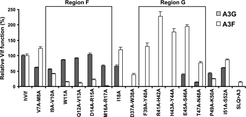
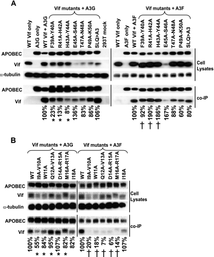
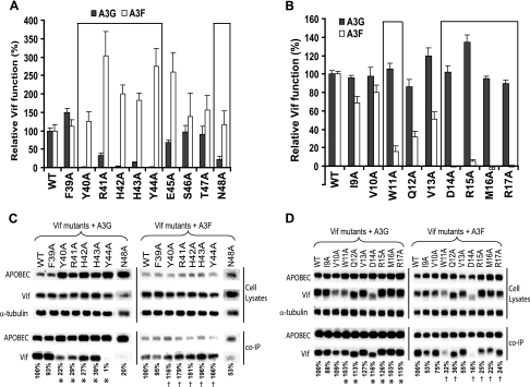
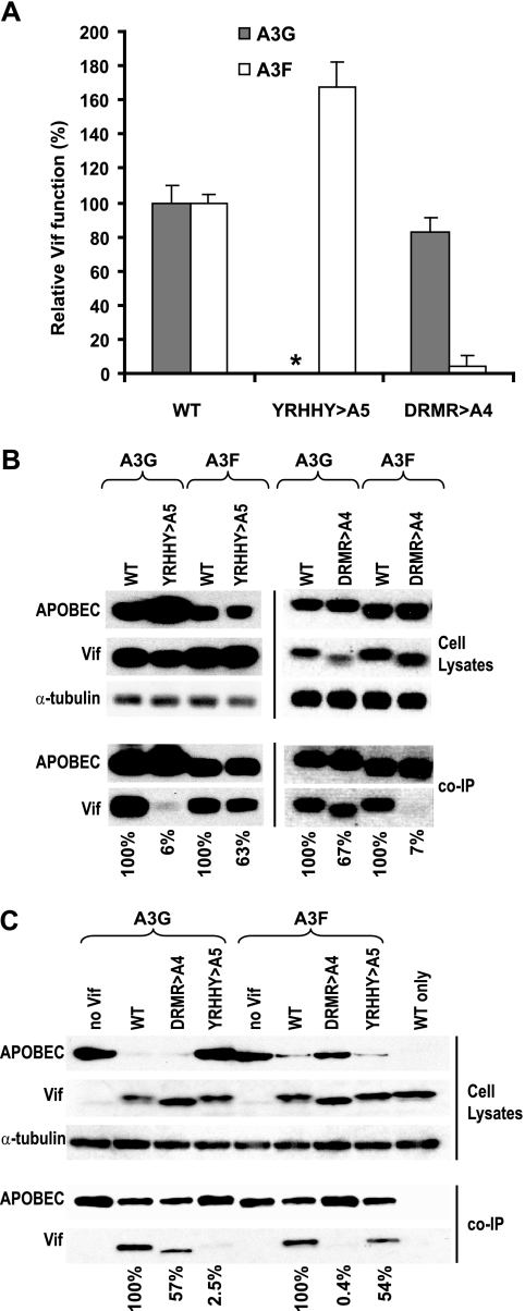
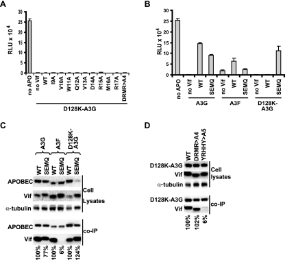
Human cytidine deaminases APOBEC3G (A3G) and APOBEC3F (A3F) inhibit replication of Vif-deficient human immunodeficiency virus type 1 (HIV-1). HIV-1 Vif overcomes these host restriction factors by binding to them and inducing their proteasomal degradation. The Vif-A3G and Vif-A3F interactions are attractive targets for antiviral drug development because inhibiting the interactions could allow the host defense mechanism to control HIV-1 replication. It was recently reported that the Vif amino acids D(14)RMR(17) are important for functional interaction and degradation of the previously identified Vif-resistant mutant of A3G (D128K-A3G). However, the Vif determinants important for functional interaction with A3G and A3F have not been fully characterized. To identify these determinants, we performed an extensive mutational analysis of HIV-1 Vif. Our analysis revealed two distinct Vif determinants, amino acids Y(40)RHHY(44) and D(14)RMR(17), which are essential for binding to A3G and A3F, respectively. Interestingly, mutation of the A3G-binding region increased Vif's ability to suppress A3F. Vif binding to D128K-A3G was also dependent on the Y(40)RHHY(44) region but not the D(14)RMR(17) region. Consistent with previous observations, subsequent neutralization of the D128K-A3G antiviral activity required substitution of Vif determinant D(14)RMR(17) with SEMQ, similar to the SERQ amino acids in simian immunodeficiency virus SIV(AGM) Vif, which is capable of neutralizing D128K-A3G. These studies are the first to clearly identify two distinct regions of Vif that are critical for independent interactions with A3G and A3F. Pharmacological interference with the Vif-A3G or Vif-A3F interactions could result in potent inhibition of HIV-1 replication by the APOBEC3 proteins.


Figures

FIG. 1.
FIG. 1.
The effect of double-alanine substitutions within Vif on its function against A3G and A3F. Double-alanine-substitution mutants were generated, and their effect on Vif function against A3G and A3F was determined. For determination of Vif mutant activity against A3G (gray bars) and A3F (white bars), 293T cells were transfected with the WT or mutant Vif expression plasmid, pHDV-EGFP, a vesicular stomatitis virus glycoprotein expression plasmid, and either an A3G-expression plasmid or an A3F-expression plasmid. The infectivity of the virus produced from the transfected cells was determined by infection of TZM-bl indicator cells and quantitation of luciferase enzyme produced in the TZM-bl cells after infection. The average relative light units (RLU) in the presence of A3G with WT Vif were 145,544 and 51,091 in the presence of A3F with WT Vif. Background levels were an average of 480 RLU. The data shown are plotted as the level of mutant Vif function as a percentage, relative to WT Vif activity (set to 100%), with standard errors of the means (SEM) from two independent experiments. Region F designates mutations in Vif that reduce activity against A3F but not A3G; region G designates mutations in Vif that reduce activity against A3G but not A3F.
FIG. 2.
FIG. 2.
Effects of single- and double-alanine-substitution mutations on binding to A3G and A3F. (A) Effects of double-alanine substitutions in region G on binding to A3G (left panel) and A3F (right panel). Either FLAG-A3G or FLAG-A3F was cotransfected into 293T cells with WT Vif- or Vif mutant-expressing plasmids. Cells were transfected with WT Vif only, A3G only, A3F only, and A3G and A3F with the SLQ Vif mutant, and mock-transfected cells served as controls. The transfected cell lysates were analyzed by Western blotting for expression of A3G or A3F (APOBEC) or Vif. The cell lysates were also analyzed for α-tubulin to control for the total amount of cell lysate analyzed. Additionally, the cell lysates were analyzed in a co-IP assay using anti-FLAG antibody to immunoprecipitate FLAG-A3G (left panel) or FLAG-A3F (right panel). The immunoprecipitated proteins were analyzed by Western blotting using anti-FLAG and anti-Vif antibodies. Band intensities were quantified from two independent experiments, and the binding efficiency of each mutant was calculated as the ratio of coimmunoprecipitated Vif to the levels of input Vif in cell lysates, normalized to the amount of immunoprecipitated A3G or A3F. The asterisks (*) and daggers (†) indicate double-alanine-substitution mutants that significantly reduced A3G binding but either enhanced or did not reduce A3F binding. (B) Effects of single- and double-alanine substitutions in region F on binding to A3G (left panel) and A3F (right panel). Either FLAG-A3G or FLAG-A3F was cotransfected into 293T cells with WT Vif- or Vif mutant-expressing plasmids. Cells transfected with WT Vif only served as a control. The transfected cell lysates were analyzed for expression of FLAG-A3G, FLAG-A3F, Vif, and α-tubulin as described above. The cell lysates were also analyzed in co-IP assays, the band intensities from two independent experiments were quantified, and the binding efficiency of each mutant was calculated as described above. The asterisks (*) and daggers (†) indicate double-alanine-substitution mutants that significantly reduced A3F binding (right panel) but did not influence A3G binding (left panel).
FIG. 3.
FIG. 3.
The effects of single-alanine-substitution mutations on Vif function and binding to APOBEC3 proteins. (A) The effects of single-alanine substitutions in region G on Vif's function against A3G and A3F. The function of Vif against A3G and A3F was determined using the assay described in the Fig. 1 legend. Single-alanine substitutions of Y40 to Y44 and N48 significantly reduced Vif function against A3G but either enhanced or did not influence Vif function against A3F. The average RLU in the presence of A3G with WT Vif were 200,550 and 130,886 in the presence of A3F with WT Vif. Background levels were an average of 350 RLU. The results are presented as a percentage of WT Vif binding with SEM from two independent experiments. (B) The effect of single-alanine substitutions in region F on Vif function against A3G and A3F. Substitutions of W11 or D14 to R17 significantly reduced Vif function against A3F but not A3G. The average RLU in the presence of A3G with WT Vif were 117,796 and 64,457 in the presence of A3F with WT Vif. Background levels were an average of 430 RLU. The results are presented as a percentage of WT Vif binding with SEM from two independent experiments. (C) Effects of single-alanine substitutions in region G on binding to A3G (left panel) and A3F (right panel). Western blotting analysis and co-IP assays were performed as described in Fig. 2 legend. The asterisks (*) and daggers (†) indicate single-alanine-substitution mutants that significantly reduced A3G binding (left panel) but either enhanced or did not influence A3F binding (left panel). (D) Effects of single-alanine substitutions in region F on binding to A3G (left panel) or A3F (right panel). Western blotting analysis and co-IP assays were performed as described in the Fig. 2 legend. The asterisks (*) and daggers (†) indicate single-alanine-substitution mutants that significantly reduced A3F binding (right panel) but did not significantly influence A3G binding (left panel). A representative analysis is shown, and the results from two independent experiments are presented as a percentage of WT Vif binding.
FIG. 4.
FIG. 4.
Effects of multiple-alanine substitutions in regions G and F on Vif function and APOBEC3 binding. (A) Amino acids Y40 to Y44 in region G were replaced by alanines to create YRHHY>A5; amino acids D14 to R17 in region F were replaced by alanines to generate DRMR>A4. The effects of these mutations on Vif function were determined as described in the Fig. 1 legend. The asterisk denotes that the effect of YRHHY>A5 mutation on function against A3G was tested, but the bar graph and the error bars are too small to be seen. The average RLU in the presence of A3G with WT Vif were 78,964 and 45,260 in the presence of A3F with WT Vif. Background levels were an average of 265 RLU. The results are presented as a percentage of WT Vif binding with SEM from two independent experiments. (B) Effects of multiple-alanine substitutions on APOBEC3 binding at a 1:1 molar ratio. The effects of the YRHHY>A5 and the DRMR>A4 mutations on A3G and A3F binding at a 1:1 molar ratio of APOBEC3:Vif are shown in the left and right panels, respectively. Western blotting analysis and co-IP assays were performed as described in the Fig. 2 legend. A representative analysis is shown, and the results from two independent experiments are presented as a percentage of WT Vif binding. (C) Effects of multiple-alanine substitutions on APOBEC3 binding at a 1:5 molar APOBEC3:Vif ratio. The effects of the YRHHY>A5 and the DRMR>A4 mutations on A3G and A3F binding at a 1:5 molar ratio of APOBEC3:Vif are shown. Western blotting analysis and co-IP assays were performed as described in the Fig. 2 legend.
FIG. 5.
FIG. 5.
Effects of single- and multiple-substitution mutations on Vif function and binding to A3G, A3F, and D128K-A3G. (A) Effect of the single-alanine mutants in region F on Vif function against D128K-A3G. The effects of these mutations on Vif function against D128K-A3G were determined as described in the Fig. 1 legend. The results shown are the RLU and SEM from two independent experiments. (B) Effect of the DRMR>SEMQ substitution on Vif function. The effects of the DRMR>SEMQ mutation on Vif function against A3G, A3F, and D128K-A3G were determined as described in the Fig. 1 legend. The results shown are RLU and SEM from three independent experiments. (C) Effect of the DRMR>SEMQ mutation on binding to A3G, A3F, and D128K-A3G. Western blotting analysis of cell lysates, co-IP assays, quantitation of band intensities, and calculation of APOBEC3 binding efficiencies were performed as described in the Fig. 2 legend. A representative blot is shown. The results from two independent experiments are presented as percentages of WT Vif binding. (D) Effect of the DRMR>A4 and YRHHY>A5 mutations on Vif binding to D128K-A3G. Western blotting analysis of cell lysates, co-IP assays, quantitation of band intensities, and calculation of D128K-A3G binding efficiencies were performed as described in the Fig. 2 legend. A representative blot is shown, and the results from two independent experiments are presented as a percentage of WT Vif binding.

Similar articles

Cited by

References

    1. Bogerd, H. P., B. P. Doehle, H. L. Wiegand, and B. R. Cullen. 2004. A single amino acid difference in the host APOBEC3G protein controls the primate species specificity of HIV type 1 virion infectivity factor. Proc. Natl. Acad. Sci. USA 101:3770-3774. - PMC - PubMed
    1. Conticello, S. G., R. S. Harris, and M. S. Neuberger. 2003. The Vif protein of HIV triggers degradation of the human antiretroviral DNA deaminase APOBEC3G. Curr. Biol. 13:2009-2013. - PubMed
    1. Derdeyn, C. A., J. M. Decker, J. N. Sfakianos, X. Wu, W. A. O'Brien, L. Ratner, J. C. Kappes, G. M. Shaw, and E. Hunter. 2000. Sensitivity of human immunodeficiency virus type 1 to the fusion inhibitor T-20 is modulated by coreceptor specificity defined by the V3 loop of gp120. J. Virol. 74:8358-8367. - PMC - PubMed
    1. Douaisi, M., S. Dussart, M. Courcoul, G. Bessou, E. C. Lerner, E. Decroly, and R. Vigne. 2005. The tyrosine kinases Fyn and Hck favor the recruitment of tyrosine-phosphorylated APOBEC3G into vif-defective HIV-1 particles. Biochem. Biophys. Res. Commun. 329:917-924. - PubMed
    1. Fujita, M., A. Sakurai, A. Yoshida, M. Miyaura, A. H. Koyama, K. Sakai, and A. Adachi. 2003. Amino acid residues 88 and 89 in the central hydrophilic region of human immunodeficiency virus type 1 Vif are critical for viral infectivity by enhancing the steady-state expression of Vif. J. Virol. 77:1626-1632. - PMC - PubMed

Publication types

MeSH terms

LinkOut - more resources