

Human parainfluenza viruses hPIV1 and hPIV3 bind oligosaccharides with alpha2-3-linked sialic acids that are distinct from those bound by H5 avian influenza virus hemagglutinin
- PMID: 17522226
- PMCID: PMC1951310
- DOI: 10.1128/JVI.00718-07
Human parainfluenza viruses hPIV1 and hPIV3 bind oligosaccharides with alpha2-3-linked sialic acids that are distinct from those bound by H5 avian influenza virus hemagglutinin
Abstract
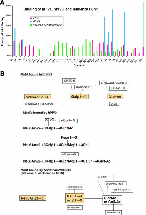
We investigated the binding of human parainfluenza virus types 1 and 3 (hPIV1 and hPIV3, respectively) to the glycan array of the Consortium for Functional Glycomics and binding and their release from erythrocytes under conditions where neuraminidase is inactive or active. hPIV1 and hPIV3 bind modifications of Neu5Acalpha2-3Galbeta1-4GlcNAc, including the sialyl-Lewis(x) motif and structures containing 6-sulfogalactose. hPIV1 and hPIV3 thus bind typical N-linked glycans, in contrast to avian influenza virus H5 hemagglutinin (J. Stevens, O. Blixt, T. M. Tumpey, J. K. Taubenberger, J. C. Paulson, and I. A. Wilson, Science 312:404-410, 2006), which binds less-common motifs. While the receptor is not the sole determinant of tropism, hPIV or H5 influenza virus infection of specific cells that express receptors may contribute to their different pathologies.
Figures

References
-
- Ah-Tye, C., S. Schwartz, K. Huberman, E. Carlin, and A. Moscona. 1999. Virus-receptor interactions of human parainfluenza viruses types 1, 2 and 3. Microb. Pathog. 27:329-336. - PubMed
-
- Corfield, A. P., M. Wember, R. Schauer, and R. Rott. 1982. The specificity of viral sialidases. The use of oligosaccharide substrates to probe enzymic characteristics and strain-specific differences. Eur. J. Biochem. 124:521-525. - PubMed
-
- Gulati, U., W. Wu, S. Gulati, K. Kumari, J. L. Waner, and G. M. Air. 2005. Mismatched hemagglutinin and neuraminidase specificities in recent human H3N2 influenza viruses. Virology 339:12-20. - PubMed
Publication types
MeSH terms
Substances
Grants and funding
LinkOut - more resources
Full Text Sources
Other Literature Sources
