Comparative Study
. 2007 May 23;27(21):5535-45.
doi: 10.1523/JNEUROSCI.1139-07.2007.

Sodium valproate exerts neuroprotective effects in vivo through CREB-binding protein-dependent mechanisms but does not improve survival in an amyotrophic lateral sclerosis mouse model

Affiliations
Comparative Study

Sodium valproate exerts neuroprotective effects in vivo through CREB-binding protein-dependent mechanisms but does not improve survival in an amyotrophic lateral sclerosis mouse model

Caroline Rouaux et al. J Neurosci. .

Abstract

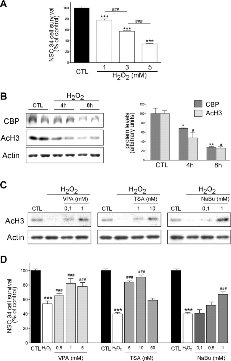
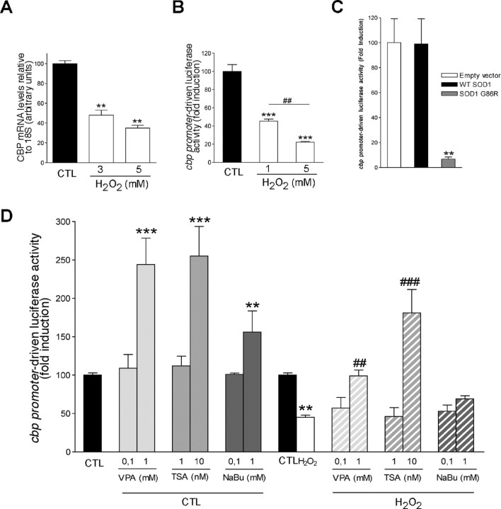
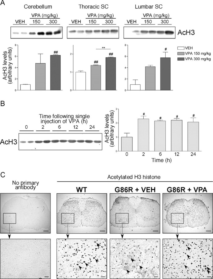
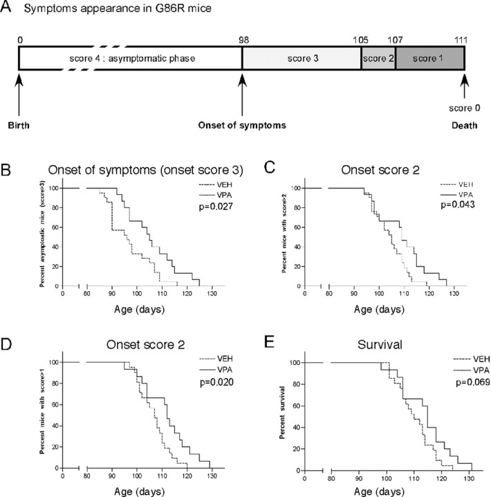
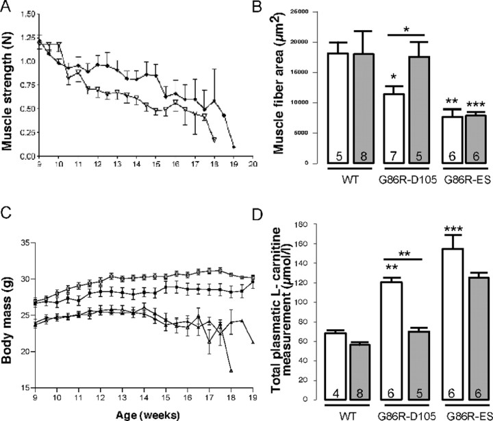
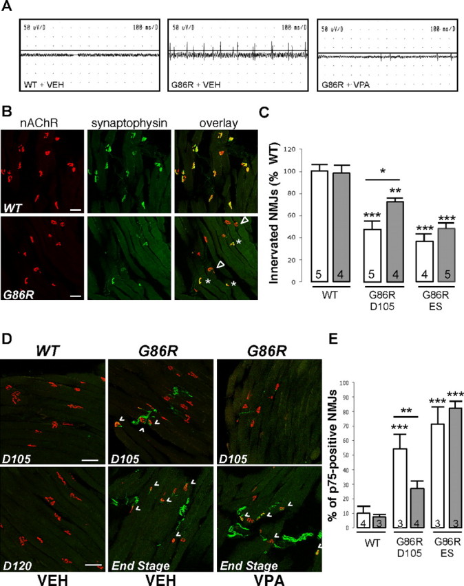
Amyotrophic lateral sclerosis (ALS) is characterized by motoneuron (MN) degeneration, generalized weakness, and muscle atrophy. The premature death of MNs is thought to be a determinant in the onset of this disease. In a transgenic mouse model of ALS expressing the G86R mutant superoxide dismutase 1 (mSOD1), we demonstrated previously that CREB (cAMP response element-binding protein)-binding protein (CBP) and histone acetylation levels were specifically decreased in nuclei of degenerating MNs. We show here that oxidative stress and mSOD1 overexpression can both impinge on CBP levels by transcriptional repression, in an MN-derived cell line. Histone deacetylase inhibitor (HDACi) treatment was able to reset proper acetylation levels and displayed an efficient neuroprotective capacity against oxidative stress in vitro. Interestingly, HDACi also upregulated CBP transcriptional expression in MNs. Moreover, when injected to G86R mice in vivo, the HDACi sodium valproate (VPA) maintained normal acetylation levels in the spinal cord, efficiently restored CBP levels in MNs, and significantly prevented MN death in these animals. However, despite neuroprotection, mean survival of treated animals was not significantly improved (<5%), and they died presenting the classical ALS symptoms. VPA was not able to prevent disruption of neuromuscular junctions, although it slightly delayed the onset of motor decline and retarded muscular atrophy to some extent. Together, these data show that neuroprotection can improve disease onset, but clearly provide evidence that one can uncouple MN survival from whole-animal survival and point to the neuromuscular junction perturbation as a primary event of ALS onset.


Figures

Figure 1.
Figure 1.
Oxidative stress-induced NSC34 death is accompanied by CBP and histone acetylation loss and is reversed by HDACis. A, NSC34 survival measurements performed in conditions of increasing concentrations of H2O2. ***p < 0.001 compared with control condition; ###p < 0.001 compared with precedent H2O2 concentration. B, Left, Western blot analysis of CBP and AcH3 protein levels in NSC34 cells treated or not with a pulse of 3 mm H2O2. Right, Actin was used as control for loading and served as reference for CBP and AcH3 band quantification. *p < 0.05 and **p < 0.01 compared with CBP levels in control condition; #p < 0.05 compared with AcH3 levels in control condition. C, Western blot analysis of AcH3 levels in NSC34 cells treated or not with a pulse of 3 mm H2O2, in presence or absence of increasing concentrations of HDACis (VPA, TSA, or NaBu). Actin served as gel loading control. D, NSC34 survival measurements in presence or absence of increasing concentrations of HDACis (VPA, TSA, or NaBu). ***p < 0.001 compared with control condition; ###p < 0.001 compared with H2O2 condition. CTL, Control.
Figure 2.
Figure 2.
Oxidative stress-induced CBP loss results from transcriptional repression that can be reversed by HDACis. A, cbp expression level was assessed by Q-PCR in NSC34 cells maintained in control conditions (CTL) or treated for 8 h with increasing concentrations of H2O2. B, cbp promoter-driven activity in control or oxidative stress (H2O2) conditions. C, NSC34 cells were transiently cotransfected with cbp promoter-luciferase reporter, together with the empty vector pRC-CMV, or vectors encoding either the WT or the mutated SOD1(G86R) protein. D, Effects of HDACis (VPA, TSA, or NaBu) on cbp promoter-driven activity in NSC34 cells under control (solid bars) or oxidative stress (hatched bars). **/##p < 0.01 and ***/###p < 0.001 when compared with control (*) or H2O2 (#) conditions (A, B, D) or to WT SOD1 condition (C).
Figure 3.
Figure 3.
Single and chronic VPA injections increase histone acetylation in nervous tissues in vivo. A, Six 90-d-old WT mice received a single injection of VEH (0.09% NaCl) or 150 or 300 mg/kg VPA. Twenty-four hours later, cerebella and spinal cords (SC) were carefully dissected and processed for acid extraction of histones. Top, Five micrograms of purified histones were then analyzed by Western blot to detect specific AcH3. Bottom, Bands were quantified. #p < 0.05 and ##p < 0.01 compared with VEH condition; **p < 0.01. B, WT mice were injected once with a dose of 250 mg/kg VPA and killed 0, 2, 6, 12, or 24 h later. Left, Spinal cords were dissected and processed for Western blot analysis. Right, Bands were quantified. #p < 0.05 compared with time 0. C, Spinal cord sections from WT (n = 5) or end-stage G86R mice daily injected with VEH (n = 5) or 250 mg/kg VPA (n = 5) were taken, and sections were immunostained using an anti-AcH3 antibody followed by a peroxidase revelation. Representative pictures are shown. Arrowheads depict lumbar motor neurons. Scale bars: top, 200 μm; bottom, 40 μm.
Figure 4.
Figure 4.
Chronic VPA administration delays motoneuronal death, maintains high levels of CBP protein, and upregulates CREB/CBP-dependent transcription in vivo. WT and G86R mice were chronically injected with VEH (0.09% NaCl) or VPA (250 mg · kg−1 · d−1) from 60 d of age until score 2 (105 d; G86R-D105) or score 0 (end stage; G86R-ES). A, Nonadjacent spinal cord sections from WT (n = 5) or transgenic mice were immunostained using an anti-ChAT antibody followed by a FITC-conjugated antibody. ChAT-positive MNs with an area >600 μm2 are represented as means ± SEM per section. The counting was performed on G86R-D105 and G86R-ES; white and gray histograms correspond to untreated and VPA-treated animals, respectively. *p < 0.05 and ***p < 0.0001 compared with WT conditions; #p < 0.05 compared with G86R plus VEH condition. B, Representative semithin section of an L4 ventral root from a WT mouse or a G86R-D105. Scale bars, 50 μm. C, D, Representative pictures (C) and quantification (D) of myelinated fibers occupancy in the different groups of mice (a, WT; b, G86R-D105 plus VEH; c, G86R-D105 plus VPA; d, G86R-ES plus VEH; e, G86R-ES plus VPA). Scale bar, 20 μm. D, The percentage of occupancy of myelinated axons within the L4 VR is represented for each group. The data represent means ± SEM. The number of each animal tested is noted within the histogram. Statistical analysis were performed with ANOVA followed by the post hoc Newman–Keuls multiple-comparisons test (Prism). All situations are compared with WT, and in each group, VPA treatment is compared with VEH. *p < 0.05; **p < 0.001. E, Representative pictures of CBP protein immunostaining in lumbar spinal cord from WT and G86R mice (score 0) chronically injected with VEH or VPA (250 mg · kg−1 · d−1). Arrowheads depict lumbar motor neurons. Scale bars, 40 μm. F, Expression of bcl-2 and smn were analyzed by semiquantitative RT-PCR 24 h after injection in spinal cord extracts from 105-d-old WT or G86R (score 2) that received or not an acute injection of VEH, VPA (250 mg/kg), TSA (2 mg/kg), or NaBu (640 mg/kg). Agarose gels were scanned, and specific bands were quantified. The results are represented relative to the expression levels of the house-keeping 18S gene mRNA levels. #p < 0.05 and ##p < 0.001 compared with the untreated G86R condition.
Figure 5.
Figure 5.
Chronic VPA delays ALS onset but does not improve mouse survival. A, Time course of clinical rating scale established on a cohort of G86R mice and representing the mean age of appearance of symptoms (see Material and Methods). B–E, G86R mice were chronically injected with VEH (0.09% NaCl; n = 21) or VPA (250 mg · kg−1 · d−1; n = 15) from 60 d of age (score 4) until the end of their life (score 0). B–D, Kaplan–Meier curves representing the proportion of G86R mice with a score >3 (B), >2 (C), and >1 (D). E, Kaplan–Meier curve representing the survival of G86R mice relative to time and treatment.
Figure 6.
Figure 6.
Chronic VPA administration delays but does not prevent loss of muscle function and does not improve body mass. WT and G86R mice were chronically injected with VEH (0.09% NaCl) or VPA (250 mg · kg−1 · d−1) from 60 d of age until score 2 (105 d; D105) or score 0 (end stage). A, Evolution of muscle strength assessed by grip test twice a week. ▿, G86R (n = 21); ♦, G86R plus VPA (n = 15). B, Muscle fiber area from WT or G86R at D105 and end stage was measured (in square micrometers) on transversal cross sections of gastrocnemius muscle (10 μm thick). C, Evolution of body weight during the survival experiment. □, WT (n = 8); ■, WT plus VPA (n = 10); ▵, G86R (n = 21); ▴, G86R plus VPA (n = 15). D, Concentration of total plasmatic l-carnitine was measured in the different groups of animals at D105 and end stage. Results are given in micromoles per liter. B, D, White and gray histograms correspond to untreated and VPA-treated animals, respectively. The data represent means ± SEM. The number of each animal tested is noted within the histogram. Statistical analysis was performed with ANOVA followed by the post hoc Newman–Keuls multiple-comparisons test (Prism). All situations are compared with WT, and in each group, VPA treatment (gray histograms) is compared with VEH (white histograms). *p < 0.05; **p < 0.01; ***p < 0.001.
Figure 7.
Figure 7.
Chronic VPA delays but does not prevent skeletal muscle denervation. A, Representative electromyography recordings performed on 100-d-old WT mice (n = 4) or G86R mice (score 3) daily injected with VEH (n = 3) or VPA (n = 3). B, Typical confocal photographs representing innervated NMJs in the gastrocnemius muscle of a WT and a G86R (end-stage) mouse. nAChRs are labeled with fluorescent α-BGT binding (red), and nerve terminals are labeled by immunochemistry of synaptophysin (green). Fully and partially innervated nAChR clusters are denoted by asterisks and arrowheads, respectively, Scale bars, 25 μm. C, Count of innervated NMJs in each experimental group. D, Visualization of nAChR (α-BGT; red) and p75 immunoreactivity (green) in WT and mutant G86R mice chronically treated with VPA or not as noted. A representative overlay (confocal analysis) is shown for each animal group. NMJs counted as positive are shown with arrowheads. WT mice corresponding to each disease stage tested were killed as noted (D105 and D120). Scale bars, 25 μm. E, Counts of p75-positive NMJs in the gastrocnemius muscle of WT and G86R chronically treated with vehicle or VPA. C, E, The data represent means ± SEM. The number of each animal tested is noted within the histogram. Statistical analysis were performed with ANOVA followed by the post hoc Newman–Keuls multiple-comparisons test (Prism) All situations are compared with WT, and in each group, VPA treatment (gray histograms) is compared with VEH (white histograms). *p < 0.05; **p < 0.01; ***p < 0.001.

References

    1. Andersen PM, Sims KB, Xin WW, Kiely R, O'Neill G, Ravits J, Pioro E, Harati Y, Brower RD, Levine JS, Heinicke HU, Seltzer W, Boss M, Brown RH., Jr Sixteen novel mutations in the Cu/Zn superoxide dismutase gene in amyotrophic lateral sclerosis: a decade of discoveries, defects and disputes. Amyotroph Lateral Scler Other Motor Neuron Disord. 2003;4:62–73. - PubMed
    1. Bannister AJ, Kouzarides T. The CBP co-activator is a histone acetyltransferase. Nature. 1996;384:641–643. - PubMed
    1. Bensimon G, Lacomblez L, Meininger V. A controlled trial of riluzole in amyotrophic lateral sclerosis. ALS/Riluzole Study Group. N Engl J Med. 1994;330:585–591. - PubMed
    1. Bjornskov EK, Norris FH, Jr, Mower-Kuby J. Quantitative axon terminal and end-plate morphology in amyotrophic lateral sclerosis. Arch Neurol. 1984;41:527–530. - PubMed
    1. Boillee S, Yamanaka K, Lobsiger CS, Copeland NG, Jenkins NA, Kassiotis G, Kollias G, Cleveland DW. Onset and progression in inherited ALS determined by motor neurons and microglia. Science. 2006;312:1389–1392. - PubMed

Publication types

MeSH terms