

Incidence of gliomas by anatomic location
- PMID: 17522333
- PMCID: PMC1907421
- DOI: 10.1215/15228517-2007-016
Incidence of gliomas by anatomic location
Abstract
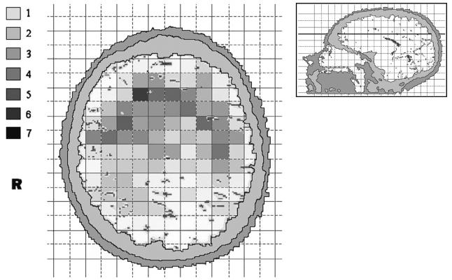
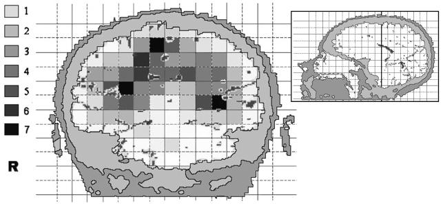
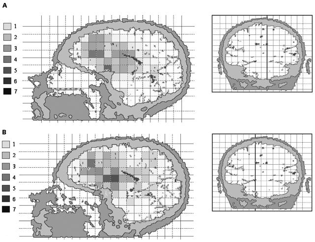
The anatomic location of a glioma influences prognosis and treatment options. The aim of our study was to describe the distribution of gliomas in different anatomic areas of the brain. A representative population-based sample of 331 adults with glioma was used for preliminary analyses. The anatomic locations for 89 patients from a single center were analyzed in more detail from radiologic imaging and recorded on a three-dimensional 1 x 1 x 1-cm grid. The age-standardized incidence rate of gliomas was 4.7 per 100,000 person-years. The most frequent subtypes were glioblastoma (47%) and grade II-III astrocytoma (23%), followed by oligodendroglioma and mixed glioma. The gliomas were located in the frontal lobe in 40% of the cases, temporal in 29%, parietal in 14%, and occipital lobe in 3%, with 14% in the deeper structures. The difference in distribution between lobes remained after adjustment for their tissue volume: the tumor:volume ratio was 4.5 for frontal, 4.8 for temporal, and 2.3 for parietal relative to the occipital lobe. The area with the densest occurrence was the anterior subcortical brain. Statistically significant spatial clustering was found in the three-dimensional analysis. No differences in location were found among glioblastoma, diffuse astrocytoma, and oligodendroglioma. Our results demonstrate considerable heterogeneity in the anatomic distribution of gliomas within the brain.
Figures
References
-
- Tola MR, Casetta I, Granieri E, et al. Intracranial gliomas in Ferrara, Italy, 1976 to 1991. Acta Neurol Scand. 1994;90:312–317. - PubMed
-
- Inskip P, Linet MS, Heineman EF. Etiology of brain tumors in adults. Epidemiol Rev. 1995;17:382–414. - PubMed
-
- Legler J, Gloecker Ries LA, Smith MA, et al. Brain and other central nervous system cancers: recent trends in incidence and mortality. J Natl Cancer Inst. 1999;91:1382–1390. - PubMed
-
- Lönn S, Klaeboe L, Mathiesen T, et al. Incidence trends of adult primary intracerebral tumors in four Nordic countries. Int J Cancer. 2004;108:450–455. - PubMed
-
- Hess KR, Broglio KR, Bondy ML. Adult glioma incidence trends in the United States 1977–2000. Cancer. 2004;101:2293–2299. - PubMed
Publication types
MeSH terms
LinkOut - more resources
Full Text Sources
Other Literature Sources
Medical
