

Detection of simulated microcalcifications in a phantom with digital mammography: effect of pixel size
- PMID: 17522348
- PMCID: PMC2430729
- DOI: 10.1148/radiol.2441060977
Detection of simulated microcalcifications in a phantom with digital mammography: effect of pixel size
Abstract
Purpose: To evaluate the effect of pixel size on the detection of simulated microcalcifications in a phantom with digital mammography.
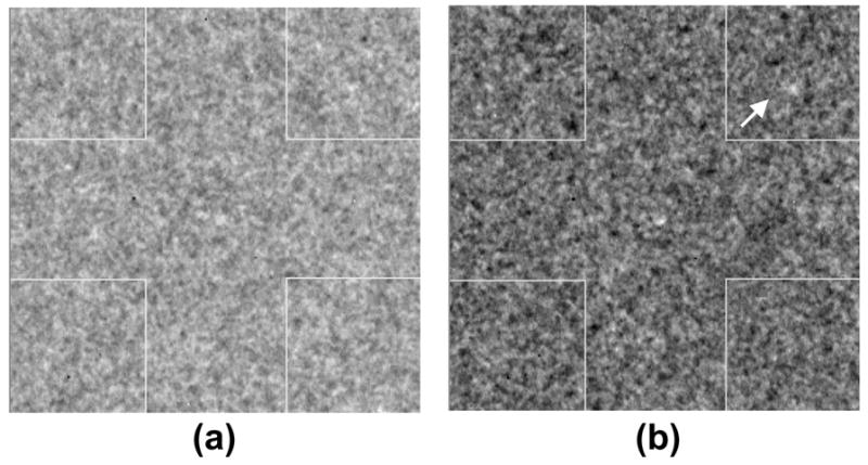
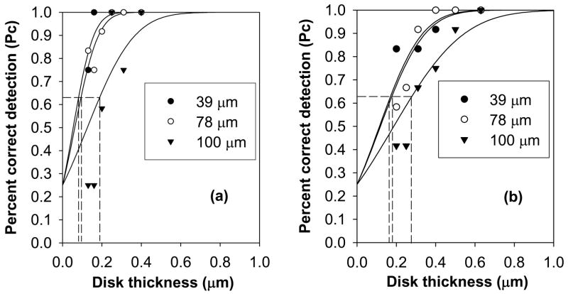
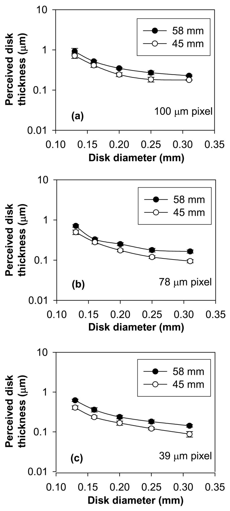
Materials and methods: A high-spatial-resolution prototype imager that yields variable pixel size (39 and 78 microm) and a clinical full-field digital mammography (FFDM) system that yields a 100-microm pixel size were used. Radiographic images of a contrast-detail (CD) phantom were obtained to perform four-alternative forced-choice observer experiments. Polymethylmethacrylate was added to obtain phantom thicknesses of 45 and 58 mm, which are typical breast thicknesses encountered in mammography. Phantom images were acquired with both systems under nearly identical exposure conditions by using an antiscatter grid. Twelve images were acquired for each phantom thickness and pixel size (for a total of 72 images), and six observers participated in this study. Observer responses were used to compute the fraction of correctly detected disks. A signal detection model was used to fit the recorded data from which CD characteristics were obtained. Repeated-measures analyses with mixed-effects linear models were performed for each of the six observers. All statistical tests were two sided and unadjusted for multiple comparisons. A P value of .05 or less was considered to indicate a significant difference.
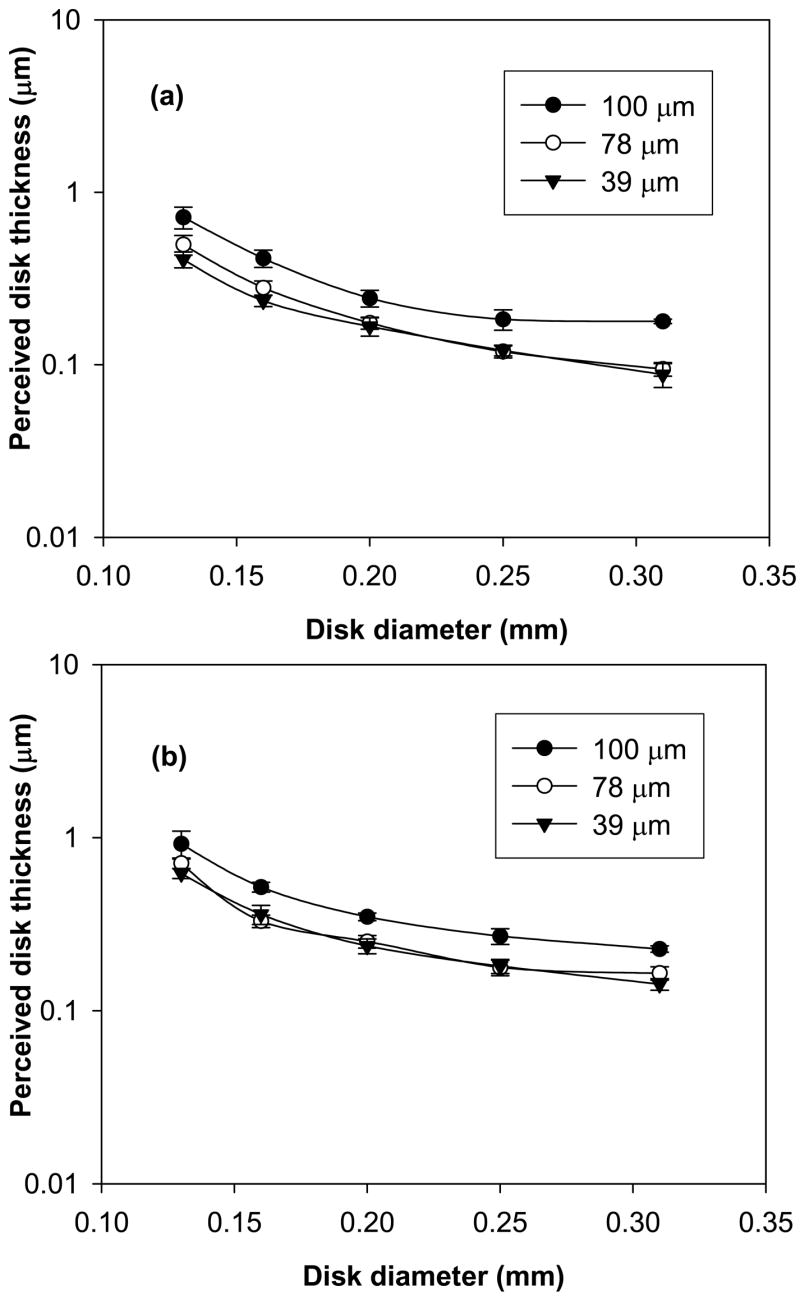
Results: Statistical analysis revealed significantly better CD characteristics with 39- and 78-microm pixel sizes compared with 100-microm pixel size for all disk diameters and phantom thicknesses (P<.001). Increase in phantom thickness degraded CD characteristics regardless of pixel size (P<.001).
Conclusion: On the basis of the conditions of this study, reducing pixel size below 100 mum with low imaging system noise enhances the visual perception of small objects that correspond to typical microcalcifications.
(c) RSNA, 2007.
Figures
References
-
- Pisano ED, Gatsonis C, Hendrick E, Yaffe M, Baum JK, Acharyya S, Conant EF, Fajardo LL, Bassett L, D’Orsi CJ, Jong R, Rebner M. Diagnostic performance of digital versus film mammography for breast-cancer screening. N Engl J Med. 2005;353:1773–1783. - PubMed
-
- Ikeda DM, Andersson I. Ductal carcinoma in situ: atypical mammographic appearances. Radiology. 1989;172:661–666. - PubMed
-
- Stomper PC, Connolly JL, Meyer JE, Harris JR. Clinically occult ductal carcinoma in situ detected with mammography: analysis of 100 cases with radiologic-pathologic correlation. Radiology. 1989;172:235–241. - PubMed
-
- Stomper PC, Connolly JL. Mammographic features predicting an extensive intraductal component in early-stage infiltrating ductal carcinoma. American Journal of Roentgenology. 1992;158:269–272. - PubMed
-
- Kinkel K, Gilles R, Feger C, Guinebretiere JM, Tardivon AA, Masselot J, Vanel D. Focal areas of increased opacity in ductal carcinoma in situ of the comedo type: mammographic-pathologic correlation. Radiology. 1994;192:443–446. - PubMed
Publication types
MeSH terms
Grants and funding
LinkOut - more resources
Full Text Sources
Medical
