Review
. 2007 Jun;7(6):429-40.
doi: 10.1038/nrc2148.

The cofilin pathway in breast cancer invasion and metastasis

Affiliations
Review

The cofilin pathway in breast cancer invasion and metastasis

Weigang Wang et al. Nat Rev Cancer. 2007 Jun.

Abstract

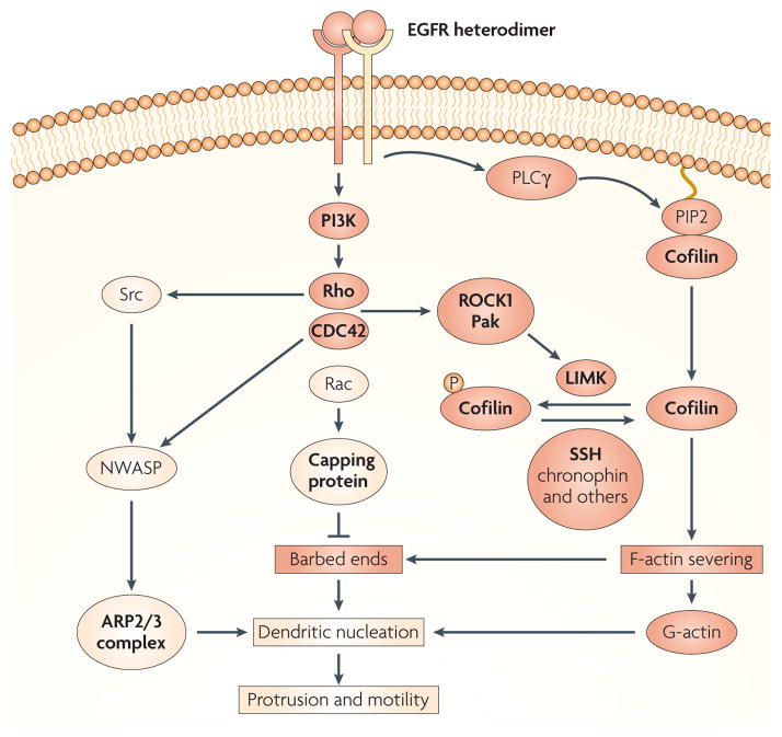
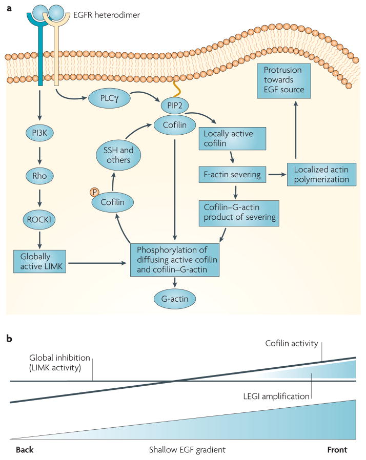
Recent evidence indicates that metastatic capacity is an inherent feature of breast tumours and not a rare, late acquired event. This has led to new models of metastasis. The interpretation of expression-profiling data in the context of these new models has identified the cofilin pathway as a major determinant of metastasis. Recent studies indicate that the overall activity of the cofilin pathway, and not that of any single gene within the pathway, determines the invasive and metastatic phenotype of tumour cells. These results predict that inhibitors directed at the output of the cofilin pathway will have therapeutic benefit in combating metastasis.


Conflict of interest statement

Competing interests statement

The authors declare no competing financial interests.

Figures

Figure 1
Figure 1. The EGF-regulated cofilin pathway
The cofilin pathway is activated in tumour cells by stimuli in the microenvironment, such as epidermal growht factor (EGF), detected by the EGF receptor (EGFR) and an unidentified subunit of the ErbB family, which could be either ERBB2 or ERBB3. In animal models ERBB2 has been shown to have a large potentiating effect on EGF-stimulated protrusion and cell migration in mammary tumours. EGFR heterodimers, with either ERBB2 or ERBB3, can activate phosphatidylinositol 3-kinase (PI3K) and then a group of small G-proteins (Rho, CDC42 (cell division cycle 42) and Rac) and their cofilin-regulating kinases (ROCK1 and Pak). These kinases stimulate LIM kinase (LIMK) to phosphorylate cofilin (P-cofilin), thereby inactivating it. And phosphatases such as slingshot (SSH), chronophin, type 1, 2A and 2B phosphatases, can dephosphorylate cofilin upon activation making it potentially active (if not bound to phosphatidylinositol-4,5-bisphosphate (PIP2) and if the pH is above 7.0). Although SSH can be activated by F-actin binding and phosphorylation by PAK4 in vitro, the mechanism for chronophin activation, is currently unknown. In addition, EGFR–ERBB2 can activate phospholipase Cγ (PLCγ) which is proposed to activate cofilin by the hydrolysis of PIP2 (REF. 29), thereby releasing cofilin from the cell membrane. Cofilin activated by either path severs mother filaments (older pre-existing filaments) to produce free barbed ends leading to the elongation of newly polymerized actin filaments that are preferred for dendritic nucleation by the ARP2/3 complex (BOX 1) and G-actin resulting from the depolymerization of pointed ends produced by the same severing reaction. In invasive tumour cells, subunits of the ARP2/3 complex are coordinately overexpressed along with genes of the cofilin pathway,, thereby potentially increasing the synergistic interaction between the cofilin and ARP2/3 complex to cause dendritic nucleation, which pushes on the cell membrane to cause cell protrusion,. The ARP2/3 complex is activated by members of the WASP (Wiskott-Aldrich syndrome protein) family, including WAVE2 and NWASP. NWASP is known to be activated in invadopodia, under the regulation of CDC42 and Rho and/or Src-mediated phosphorylation. The cofilin pathway is coordinately regulated in invasive tumour cells during cell migration in vivo (genes that are upregulated in rat mammary tumours are highlighted in bold), suggesting that the cofilin pathway (which is shown in red boxes and circles) has a direct role in determining the invasive and metastatic phenotype.
Figure 2
Figure 2. The spatial and temporal localization of cofilin activity in response to EGF stimulation
a | In vivo studies in tumour cells indicate that EGF-stimulated phospholipase Cγ (PLCγ) hydrolyses phosphatidylinositol-4,5-bisphosphate (PIP2), causing the release of active cofilin locally from its complex with PIP2 in the plasma membrane. This activates cofilin asymmetrically inside the tumour cell to generate free barbed ends adjacent to the cell membrane facing the source of epidermal growth factor (EGF). Transient cofilin activity occurs as the result of the near simultaneous activation of the molecules within the cofilin pathway that both activate cofilin (PLCγ, slingshot (SSH) and chronophin) and inhibit cofilin (LIMK) by the EGF receptor. As a result, cofilin severs filaments locally to start polymerization and cell protrusion, and global LIMK activity inactivates cofilin that diffuses from the initial site of activation, thereby spatially sharpening cofilin activity. The signalling pathways from the receptor through G-proteins and LIMK are the same as in FIG. 1 but redrawn here to show how LIMK captures cofilin that diffuses from the site of activation. This results in the sharpened localization of cofilin-dependent free barbed end production and the initiation of directional cell motility and chemotaxis. b | The cofilin activity cycle fits a local excitation global inhibition (LEGI) model of chemotaxis. Cofilin is activated asymmetrically inside the cell in response to the gradient in EGF detected by cell surface receptors from the front to the back of the cell. The asymmetry of cofilin activation inside the cell may follow the slope of the gradient of EGF. However, this shallow gradient in cofilin activation is locally sharpened by the global stimulation of LIMK activity, which is postulated to inactivate cofilin throughout the cell, resulting in a remnant of cofilin activity only on the side of the cell facing the EGF source (front). This is shown by the two lines indicating the cofilin activation gradient (sloping line) and global LIMK activity (horizontal line) resulting from the same stimulation with a shallow EGF gradient, thereby compressing the effective cofilin activity between them to sharpen cofilin activity to the side of the cell facing the EGF source.
Figure 3
Figure 3. Cofilin activity is required for the early barbed end transient responsible for the initiation of chemotaxis
a | Stimulation of mammary tumour cells with epidermal growth factor (EGF) results in two transients of barbed end formation (b), which are localized at the cell membrane. The first transient is required for initiating actin polymerization and protrusion towards the source of EGF, resulting in chemotaxis, and the second transient is required for sustaining the cell protrusions required for cell migration,. Two transients of barbed end formation have been observed in chemotactic cells (Dictyostelium, mammalian fibroblasts and macrophages) in response to various chemoattractants, indicating that this sequence of events is conserved in crawling chemotactic cells. c | In mammary tumour cells the first, but not the second, transient requires both phospholipase Cγ (PLCγ) and cofilin activity, implicating the cofilin pathway in chemotaxis (the red line shows the effect of inhibiting either PLCγ or cofilin activity),. d | Cofilin-dependent free barbed ends are sharply localized in vivo in response to a gradient of EGF. The first transient of free barbed ends (stained red) 60 seconds after the introduction of a micropipette source of EGF are found predominantly on the side of the cell facing the source of EGF (right hand image; *marks the position of EGF-filled micropipette) compared with the more uniform distribution of free barbed ends in unstimulated cells (0 seconds). Part a of the figure is taken from REF. and reproduced with permission of the Company of Biologists; parts b and c of the figure are reproduced from REF. © (2004) The Rockefeller University Press; part d is taken from REF © (2006) Elsevier Science. au, arbitrary units.

Similar articles

Cited by

References

    1. Liotta LA, Kohn EC. The microenvironment of the tumour-host interface. Nature. 2001;411:375–379. - PubMed
    1. Condeelis J, Segall JE. Intravital imaging of cell movement in tumours. Nature Rev Cancer. 2003;3:921–930. - PubMed
    1. Hanahan D, Weinberg RA. The hallmarks of cancer. Cell. 2000;100:57–70. - PubMed
    1. Mantovani A, Giavazzi R, Alessandri G, Spreafico F, Gerattini S. Characterization of tumor lines derived from spontaneous metastases of transplanted murine sarcoma. Eur J Cancer. 1981;17:71–76. - PubMed
    1. Giavazzi R, Alessandri G, Spreafico F, Garattini S, Mantovani A. Metastasizing capacity of tumour cells from spontaneous metastases of transplanted murine tumours. Br J Cancer. 1980;42:462–472. - PMC - PubMed

Publication types

Substances