Review
. 2007 Jun;210(6):642-50.
doi: 10.1111/j.1469-7580.2007.00728.x.

Use-dependent control of presynaptic calcium signalling at central synapses

Affiliations
Review

Use-dependent control of presynaptic calcium signalling at central synapses

Ricardo Scott. J Anat. 2007 Jun.

Abstract

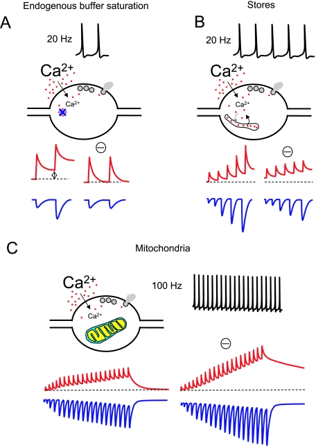
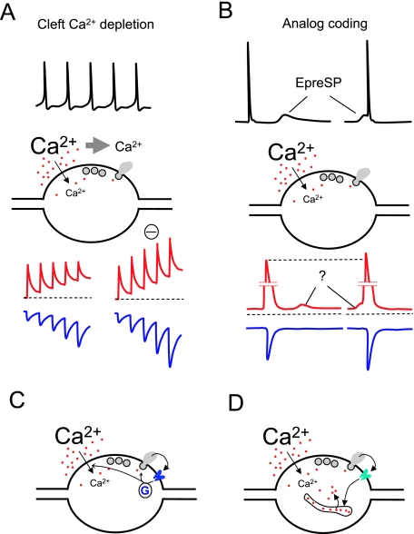
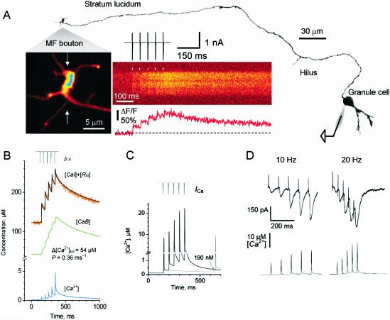
Voltage-gated Ca(2+) channels activated by action potentials evoke Ca(2+) entry into presynaptic terminals thus briefly distorting the resting Ca(2+) concentration. When this happens, a number of processes are initiated to re-establish the Ca(2+) equilibrium. During the post-spike period, the increased Ca(2+) concentration could enhance the presynaptic Ca(2+) signalling. Some of the mechanisms contributing to presynaptic Ca(2+) dynamics involve endogenous Ca(2+) buffers, Ca(2+) stores, mitochondria, the sodium-calcium exchanger, extraterminal Ca(2+) depletion and presynaptic receptors. Additionally, subthreshold presynaptic depolarization has been proposed to have an effect on release of neurotransmitters through a mechanism involving changes in resting Ca(2+). Direct evidence for the role of any of these participants in shaping the presynaptic Ca(2+) dynamics comes from direct recordings of giant presynaptic terminals and from fluorescent Ca(2+) imaging of axonal boutons. Here, some of this evidence is presented and discussed.


Figures

Fig. 1
Fig. 1
Mechanisms of control of presynaptic Ca2+ dynamics (I). For all the panels in this figure and Fig. 2 a general idealistic illustration of each of the mechanisms described in the text is shown. The stimulating protocols at which these mechanisms occur are depicted on top of each panel. Red traces represent free Ca2+ concentration. Blue traces represent postsynaptic responses. The hypothetical blockade of each mechanism is indicated with a minus symbol (‘–’). All panels represent general concepts and are not intended to show quantitative data. For an accurate idea of a realistic view of free Ca2+ dynamics and postsynaptic responses see Fig. 3. Only non-depressing synapses with a low release probability are considered. (A) The role of endogenous buffers in paired pulse facilitation (PPF) at a synaptic terminal. Two APs at 20 Hz produce two consecutive increases in Ca2+ concentration inside the terminal. The endogenous presynaptic Ca2+ buffers rapidly trap Ca2+ ions and can be partially saturated after the first AP at relatively high frequencies. Therefore, the residual Ca2+ when the second AP occurs is higher (left red trace), and this enhances transmitter release (left blue trace). The effect on free Ca2+ and EPSCs of a hypothetic blockade of buffer saturation is also illustrated. (B) Upon repetitive stimulation at 20 Hz, Ca2+ release from presynaptic Ca2+ stores can occur, contributing to the global Ca2+ signal. Through this mechanism neurotransmitter release is facilitated. The blockade of Ca2+ stores should reduce facilitation (only partially due to the presence of mechanism A) and increase resting Ca2+. (C) Intense (100 Hz) stimulation recruits presynaptic mitochondria and contributes to buffer the excess of Ca2+. In the absence of mitochondria the presynaptic Ca2+ concentration would reach higher levels during such a repetitive high-frequency stimulation, increasing the release rate.
Fig. 2
Fig. 2
Mechanisms of control of presynaptic Ca2+ dynamics (II). See also legend to Fig. 1. (A) High-frequency stimulation transiently decreases the extracellular concentration of Ca2+ in the synaptic cleft due to Ca2+ ions passing into the cells during the electrical activity. The reduction of the electrochemical gradient for Ca2+ produces a decrease of presynaptic Ca2+ entry during subsequent AP and a reduction of neurotransmitter release. A hypothetical blockade of Ca2+ depletion restablishes the amplitude of consecutive Ca2+ transients and enhances facilitation. (B) Subthreshold voltage changes in the dendrites or soma can passively travel relatively long distances along the axons and regulate neurotransmitter release when they immediately precede an AP. A suggested mechanism for this phenomenon would be that small changes in resting Ca2+ produced by such subthreshold voltage signals would add to the Ca2+ transient elicited by an AP, resulting in an enhancement of transmitter release. (C) Presynaptic metabotropic receptors can modulate Ca2+ entry and or neurotransmitter release via G-proteins. (D) Ionotropic receptors can regulate neurotransmitter release interacting with intracellular stores.
Fig. 3
Fig. 3
Ca2+ dynamics in mossy fibre giant terminals at the stratum lucidum. (A) Giant terminal (with visible filopodia) emerging from the mossy fibre of a granule cell loaded with alexa-594 and fluo-4 through a patch pipette. The morphology of the cell and the main axon is shown (collateral branches were out of focus). The cell body was briefly depolarized evoking action currents that propagated along the axon producing Ca2+-dependent fluorescence transients at the giant terminals, almost 1 mm distant. These transients were recorded by line-scanning two-photon microscopy. The average of ten fluorescent transients is shown. (B) Average response for 18 giant boutons (orange trace) in similar conditions to A. Several parameters involved in Ca2+ dynamics could be assumed (AP-dependent Ca2+ influx, calbindin concentration, dye concentration, and Kd of the dye; Scott & Rusakov, 2006). An accurate fitting of the experimental data was obtained, also rendering the kinetics of the endogenous buffer bound to Ca2+ (green trace) and the free Ca2+ concentration changes during trains of AP (blue trace). In order to constrain the unknown parameters (total Ca2+ entry and removal rate, Δ[Ca]tot and P, respectively), the experiments were repeated with fluo-4 (50 µm) and fluo-5 (200 µm). Similar values were obtained in the three conditions for these unknowns (not shown). (C) The model allowed us to extrapolate the dynamics of free Ca2+ concentration in the absence of exogenous buffers. It was also possible to simulate the free Ca2+ dynamics during different frequencies of AP. (D) Typical EPSCs recorded in CA3 pyramidal cells at 20 Hz and 50 Hz. Below, the simulations of free Ca2+ at the corresponding frequencies (modified from Scott & Rusakov, 2006).

Similar articles

Cited by

References

    1. Alle H, Geiger JR. Combined analog and action potential coding in hippocampal mossy fibers. Science. 2006;311:1290–1293. - PubMed
    1. Allen TJA, Noble D, Reuter H. Sodium–Calcium Exchange. Oxford: Oxford University Press; 1989.
    1. Awatramani GB, Price GD, Trussell LO. Modulation of transmitter release by presynaptic resting potential and background calcium levels. Neuron. 2005;48:109–121. - PubMed
    1. Billups B, Forsythe ID. Presynaptic mitochondrial calcium sequestration influences transmission at mammalian central synapses. J Neurosci. 2002;22:5840–5847. - PMC - PubMed
    1. Bischofberger J, Geiger JR, Jonas P. Timing and efficacy of Ca2+ channel activation in hippocampal mossy fiber boutons. J Neurosci. 2002;22:10593–10602. - PMC - PubMed

LinkOut - more resources