. 2007 Aug 15;313(14):3066-75.
doi: 10.1016/j.yexcr.2007.04.022. Epub 2007 Apr 27.

Cell shape regulates global histone acetylation in human mammary epithelial cells

Affiliations

Cell shape regulates global histone acetylation in human mammary epithelial cells

Johanne Le Beyec et al. Exp Cell Res. .

Abstract

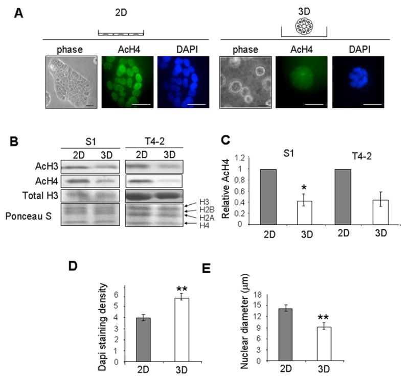
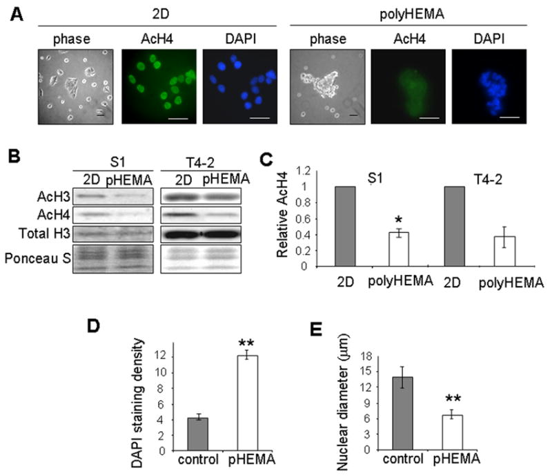
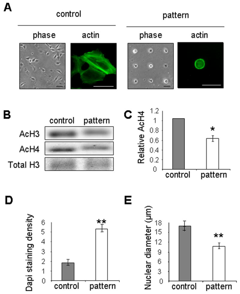
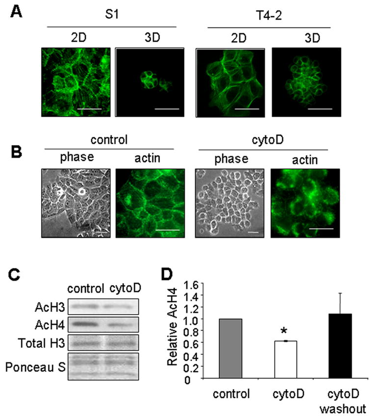
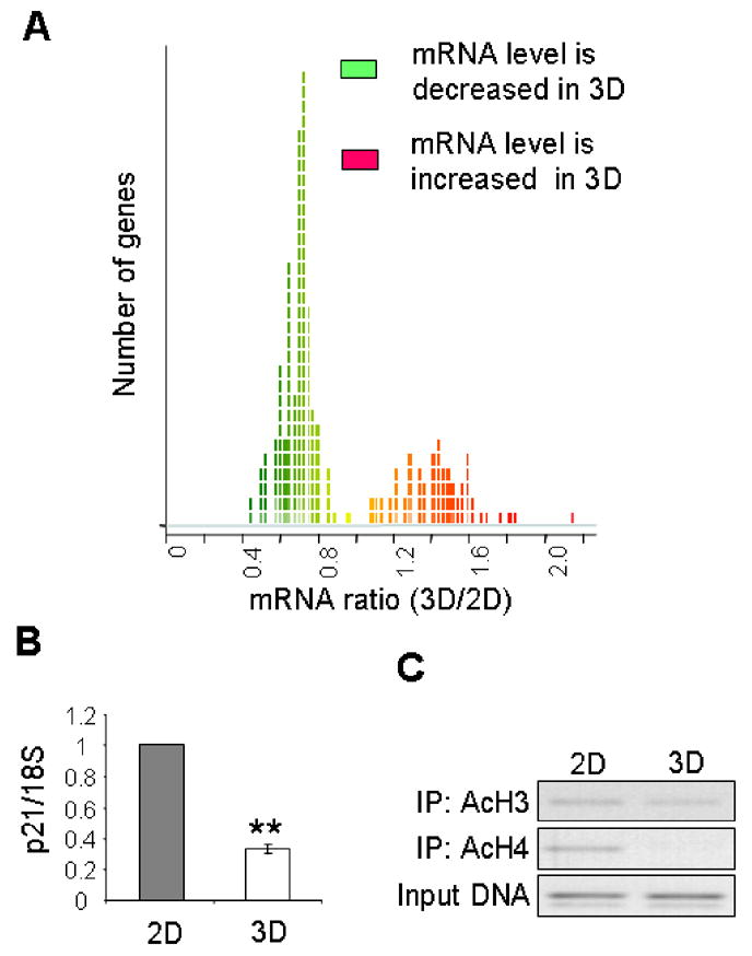
Extracellular matrix (ECM) regulates cell morphology and gene expression in vivo; these relationships are maintained in three-dimensional (3D) cultures of mammary epithelial cells. In the presence of laminin-rich ECM (lrECM), mammary epithelial cells round up and undergo global histone deacetylation, a process critical for their functional differentiation. However, it remains unclear whether lrECM-dependent cell rounding and global histone deacetylation are indeed part of a common physical-biochemical pathway. Using 3D cultures as well as nonadhesive and micropatterned substrata, here we showed that the cell 'rounding' caused by lrECM was sufficient to induce deacetylation of histones H3 and H4 in the absence of biochemical cues. Microarray and confocal analysis demonstrated that this deacetylation in 3D culture is associated with a global increase in chromatin condensation and a reduction in gene expression. Whereas cells cultured on plastic substrata formed prominent stress fibers, cells grown in 3D lrECM or on micropatterns lacked these structures. Disruption of the actin cytoskeleton with cytochalasin D phenocopied the lrECM-induced cell rounding and histone deacetylation. These results reveal a novel link between ECM-controlled cell shape and chromatin structure and suggest that this link is mediated by changes in the actin cytoskeleton.


Figures

Figure 1
Figure 1
Culturing mammary epithelial cells in 3D lrECM induces alterations in cellular morphology and global histone deacetylation. (A) Phase contrast and immunofluorescence images of AcH4 and DAPI staining in S1 cells on tissue culture plastic (2D) or within lrECM (3D). Scale bars, 50 μm; (B) Western blot analysis of AcH3 and AcH4 in S1 and T4-2 cells cultured in 2D and 3D; (C) Bar graphs quantifying the relative AcH4 levels in S1 (n=4) and T4-2 (n=2) cells cultured in 2D and 3D; error bars indicate s.e.m. (*) p<0.05; (D) Quantification of nuclear DAPI staining for S1 cells cultured in 2D and 3D. (**) p<0.01; n=20; (E) Quantification of nuclear diameter for S1 cells cultured in 2D and 3D; error bars indicate s.e.m. (**) p<0.01; n=20.
Figure 2
Figure 2
Cell rounding on the nonadhesive substratum polyHEMA induces global histone deacetylation. (A) Phase contrast images of S1 cells cultured in 2D on tissue culture plastic or rounded on polyHEMA. Immunofluorescence images of AcH4 and DAPI staining in S1 cells cultured in 2D and on polyHEMA; (B) Western blot analysis of AcH3 and AcH4 in S1 and T4-2 cells cultured in 2D or on polyHEMA; (C) Bar graphs quantifying the relative AcH4 levels in S1 (n=4) and T4-2 (n=2) cells cultured in 2D and on polyHEMA. (**) p<0.01; (D) Quantification of nuclear DAPI staining in S1 cells cultured in 2D and on polyHEMA. (**) p<0.01; n=20; (E) Quantification of nuclear diameter for S1 cells cultured in 2D and on polyHEMA. (**) p<0.01; n=20.
Figure 3
Figure 3
Cell rounding on micropatterned substrata induces histone deacetylation. (A) Phase contrast and immunofluorescence images of phalloidin-stained T4-2 cells cultured on unpatterned substratum (control) or substratum patterned with 25-μm square islands (pattern) for 24 hours. Scale bars, 50 μm; (B) Western blot analysis of AcH3 and AcH4 in T4-2 cells cultured on unpatterned and patterned substrata; (C) Bar graphs quantifying the relative AcH4 levels in T4-2 cells. (*) p<0.05, n=4; (D) Quantification of nuclear DAPI staining for T4-2 cells cultured on unpatterned and patterned substrata. (**) p<0.01; n=20; (E) Quantification of nuclear diameter for T4-2 cells cultured on unpatterned and patterned substrata. (**) p<0.01; n=20.
Figure 4
Figure 4
Actin cytoskeleton regulates cell rounding and histone deacetylation. (A) Immunofluorescence images of phalloidin-stained S1 and T4-2 cells cultured in 2D or 3D; (B) Phase contrast and immunofluorescence images of phalloidin-stained S1 cells in the presence or absence of cytochalasin D (cytoD). Scale bars, 50 μm; (C) Western blot for AcH3 and AcH4 in S1 cells in the presence or absence of cytochalasin D. (D) Bar graphs of relative AcH4 levels in the control S1 cells (control), cells treated with cytochalasin D (cytoD), and cells treated with cytochalasin D followed by removal of the drug (cytoD washout). (*) p<0.05, n=4.
Figure 5
Figure 5
Culturing mammary epithelial cells in 3D lrECM induces a global reduction in gene expression. (A) Ratio of global mRNA levels for S1 cells cultured in 2D and 3D lrECM. The x-axis shows mean ratio of 3D/2D for four experiments; all genes with p< 0.01 are displayed: 91 genes have higher mRNA levels in 3D (red); 162 genes have lower levels in 3D (green). (B) Quantitative RT/PCR analysis for p21 normalized to levels of 18S in the same samples. (**) p<0.01; (C) ChIP assay measuring levels of AcH3 and AcH4 in the p21 promoter for S1 cells cultured in 2D and 3D.

References

    1. Bissell MJ, Hall HG, Parry G. How does the extracellular matrix direct gene expression? J Theor Biol. 1982;99:31–68. - PubMed
    1. Bissell MJ, Farson D, Tung AS. Cell shape and hexose transport in normal and virus-transformed cells in culture. J Supramol Struct. 1977;6:1–12. - PubMed
    1. Folkman J, Moscona A. Role of cell shape in growth control. Nature. 1978;273:345–9. - PubMed
    1. Chen CS, Mrksich M, Huang S, Whitesides GM, Ingber DE. Geometric control of cell life and death. Science. 1997;276:1425–8. - PubMed
    1. Singhvi R, Kumar A, Lopez GP, Stephanopoulos GN, Wang DI, Whitesides GM, Ingber DE. Engineering cell shape and function. Science. 1994;264:696–8. - PubMed

Publication types