Review
. 2007 Oct;116(1):35-57.
doi: 10.1016/j.pharmthera.2007.03.004. Epub 2007 Apr 20.

Mechanisms of neurosteroid interactions with GABA(A) receptors

Affiliations
Review

Mechanisms of neurosteroid interactions with GABA(A) receptors

Gustav Akk et al. Pharmacol Ther. 2007 Oct.

Abstract

Neuroactive steroids have some of their most potent actions by augmenting the function of GABA(A) receptors. Endogenous steroid actions on GABA(A) receptors may underlie important effects on mood and behavior. Exogenous neuroactive steroids have potential as anesthetics, anticonvulsants, and neuroprotectants. We have taken multiple approaches to understand more completely the interaction of neuroactive steroids with GABA(A) receptors. We have developed many novel steroid analogues in this effort. Recent work has resulted in synthesis of new enantiomer analogue pairs, novel ligands that probe various properties of the steroid pharmacophore, fluorescent neuroactive steroid analogues, and photoaffinity labels. Using these tools, combined with receptor binding and electrophysiological assays, we have begun to untangle the complexity of steroid actions at this important class of ligand-gated ion channel.


Figures

Figure 1
Figure 1. GABAA receptor schematic and putative binding sites
A. A single subunit of the GABAA receptor, highlighting topology. M1-M4 represent transmembrane domains. The M2 transmembrane domain (gray) forms an important part of the chloride channel pore. B. Pentameric structure of a typical GABAA receptor. Several putative sites of GABA and modulatory drugs, including neurosteroids, are shown. Mutations of the β subunit affect barbiturate modulation, but no unequivocal binding site has been identified. The indication that steroids act on the GABAA receptor from within the transmembrane domains is supported by pharmacological studies and by recent site-directed mutagenesis studies (Akk et al., 2005; Hosie et al., 2006; Shu et al., 2004). C. Top-down view of the pentameric receptor showing proposed sites of potentiation and direct gating for neurosteroids, based on site-directed mutagenesis (Hosie et al., 2006).
Figure 2
Figure 2. Correlation between effects of steroid analogues on TBPS binding in rat brain membranes and loss of righting reflex (LRR) in tadpoles
This figure shows a scatter plot of data for 212 steroids and analogues. The ordinate gives the IC50 for inhibition of TBPS binding while the abscissa shows the EC50 for producing loss of righting in Xenopus tadpoles. Note that both axes are logarithmic, due to the large range of values observed (about 3.5 orders of magnitude for each parameter). The solid line shows the regression of log(IC50) on log(EC50) (slope 0.65, P = 3×10-18 that the slope does not differ from zero). The dashed line shows the line of equality. Some specific compounds are highlighted. Three pairs of enantiomers are shown, connected by thin solid lines (3α5αP, solid circles; 3α5βP, solid diamonds and ACN, solid triangles). The analogue B285 (X) has been extensively studied in single channel research, while ECN (cross) is an example of a compound with much higher potency at producing loss of righting than at inhibiting TBPS binding.
Figure 3
Figure 3. Steroid accesses receptor from membrane diffusion or from intracellular access
Cell-attached and inside-out recordings from HEK cells transfected with α1β2γ2 subunits were performed under the conditions schematized in the right panels. Downward deflections in the channel records represent channel openings. The graphs are log-binned histograms of open and closed times from the indicated patch. For all recordings, 50 μM GABA was included in the patch pipette to activate the channels. ACN (1 μM) was the steroid analogue used to potentiate channel function. Panels A-D represent cell-attached patch recordings, and panels E-G represent inside-out patch recordings. A. Patch exposed to GABA alone. The open times were 0.16 ms (49 %), 3.1 ms (37 %) and 7.8 ms (14 %). The closed times were 0.19 ms (61 %), 1.7 ms (20 %) and 21.7 ms (18 %). B. Channel activity in a patch exposed to the combination of GABA and ACN in the patch pipette. The open times were 0.27 ms (29 %), 1.4 ms (24 %) and 19.6 ms (47 %). The closed times were 0.19 ms (68 %), 1.5 ms (28 %) and 25.6 ms (5 %). Steroid increases both the duration and the amplitude of the third (longest) component of the open time distribution. Steroid also decreases the relative contribution of the third (longest) component of the closed time distribution (Akk et al., 2004). C. Channel activity in a patch indirectly exposed to ACN. Receptor function was still augmented when ACN was added to the bath solution following seal formation. The open times were 0.34 ms (33 %), 9.2 ms (34 %) and 21.3 ms (33 %). The closed times were 0.11 ms (57 %), 1.0 ms (35 %) and 25.0 ms (8 %). D. Channel activity in a patch recorded from a cell pretreated with ACN. Free bath ACN was removed before the recording was made, but potentiation of channel activity remained. The open times were 0.42 ms (19 %), 1.7 ms (38 %) and 34.5 ms (43 %). The closed times were 0.17 ms (73 %), 1.2 ms (23 %) and 22.7 ms (4 %). E. Channel activity in an excised patch taken from a cell pre-incubated with ACN. The patch was excised into a steroid-free bath, and the record was taken 1 min after excision. Augmented channel function remains despite removal of the channel from intracellular and extracellular sources of ACN. Histograms pertain to data from 0-2 min after excision. The open times were 0.31 ms (42 %), 2.1 ms (23 %) and 18.5 ms (35 %). The closed times were 0.20 ms (58 %), 1.2 ms (32 %) and 18.5 ms (9 %). F. The effect of addition of 5 mM methyl-β-cyclodextrin (CDX) to the bath to remove steroid. Cyclodextrin application immediately removed steroid-mediated potentiation. The open times were 0.46 ms (24 %), 3.4 ms (57 %) and 6.9 ms (19 %). The closed times were 0.15 ms (60 %), 1.5 ms (9 %) and 10.4 ms (31 %). G. Intracellular application of ACN results in potentiation. Channel activity in an inside-out patch exposed to 1 μM ACN applied to the cytoplasmic face of the membrane. The open times were 0.42 ms (48 %), 4.2 ms (15 %) and 23.3 ms (37 %). The closed times were 0.28 ms (48 %), 1.4 ms (42 %) and 14.5 ms (10 %). The figure is taken from data in (Akk et al., 2005). Copyright Society for Neuroscience.
Figure 4
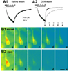
Figure 4. Effect of cyclodextrin on the washout of currents and fluorescence of a tagged steroid analogue
A1. NBD-3α5αP (300 nM) was applied to neurons in the absence of GABA, and whole-cell currents were monitored. Drug exposure is indicated by the horizontal bar. Despite rapid removal of free drug, the current decayed slowly. A2. Including 500 μM γ-cyclodextrin (CDX) in the wash following drug removal (solid trace) increased the rate of current offset. The dotted trace is a re-plot of A1. B1 and B2. Imaging of a neuron following exposure to fluorescent steroid analogue at the time points denoted by the small letters in A2. The fluorescence intensity is pseudocolored, with warm colors representing high fluorescence intensity. B1 shows loss of fluorescence with normal saline wash. B2 shows intracellular fluorescence loss is speeded by cyclodextrin wash. The figure is taken from (Akk et al., 2005). Copyright Society for Neuroscience.
Figure 5
Figure 5. Coapplication of 3α5αP with GABA affects the open time distributions
A. Modelled open time histograms for GABA (thin lines) and GABA + 1 μM 3α5αP (thick lines). The longest open time component (OT3) is shown with dashed lines. The presence of steroid results in the increase in the duration and relative frequency of OT3 (shown with arrows). The modelling was conducted using the mean values for control and steroid data from Akk et al. (2005). B. Sample single channel currents recorded in the presence of GABA and GABA + 1 μM 3α5αP. C. Concentration effect curves for the duration and fraction of OT3. The duration of OT3 increases from 5-6 ms to approximately 15 ms, and the relative frequency of OT3 increases from 0.15 to approximately 0.45 in the presence of steroid. In addition to the changes in open times, 3α5αP also affects the relative frequency of the activation-related closed time component (channel closing rate).
Figure 6
Figure 6. 17PA antagonizes 3α5αP effects on GABA receptors
A1 and A2. Comparison of the effect of 10 μM 17PA on potentiation by 0.5 μM 3α5αP (A1) and 3α5βP (A2) on GABA receptors expressed in an oocyte. The inset in panel A1 shows the structure of 17PA. GABA and drugs were co-applied. B1 and B2. Effect of 17PA (10 μM) on steroid (5 μM) activation of receptors in the absence of GABA. Note the nearly complete inhibition of direct gating by 3α5αP despite the high concentration of steroid agonist used. Note that neither potentiation nor direct activation by 3α5βP was depressed by 17PA. The data are taken from (Mennerick et al., 2004). Copyright American Society for Pharmacology and Experimental Therapeutics.
Figure 7
Figure 7. Structures of enantiomer pairs
Figure 8
Figure 8. Structures of benz[e]indene and benz[f]indenes
Figure 9
Figure 9. Structures of indicated analogues

References

    1. Adkins CE, Pillai GV, Kerby J, Bonnert TP, Haldon C, McKernan RM, Gonzalez JE, Oades K, Whiting PJ, Simpson PB. α4β3δ GABAA receptors characterized by fluorescence resonance energy transfer-derived measurements of membrane potential. J Biol Chem. 2001;276:38934–38939. - PubMed
    1. Agarwal K, Bali A, Gupta CM. Effect of phospholipid structure on stability and survival times of liposomes in circulation. Biochim Biophys Acta. 1986a;883:468–475. - PubMed
    1. Agarwal K, Bali A, Gupta CM. Influence of the phospholipid structure on the stability of liposomes in serum. Biochim Biophys Acta. 1986b;856:36–40. - PubMed
    1. Agis-Balboa RC, Pinna G, Zhubi A, Maloku E, Veldic M, Costa E, Guidotti A. Characterization of brain neurons that express enzymes mediating neurosteroid biosynthesis. Proc Natl Acad Sci U S A. 2006;103:14602–14607. - PMC - PubMed
    1. Akabas MH. GABAA receptor structure-function studies: a reexamination in light of new acetylcholine receptor structures. Int Rev Neurobiol. 2004;62:1–43. - PubMed

Publication types