. 2007 Jun 29;147(2):546-60.
doi: 10.1016/j.neuroscience.2007.02.065. Epub 2007 May 23.

Functional recovery in rats with ischemic paraplegia after spinal grafting of human spinal stem cells

Affiliations

Functional recovery in rats with ischemic paraplegia after spinal grafting of human spinal stem cells

D Cizkova et al. Neuroscience. .

Abstract

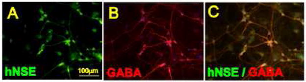
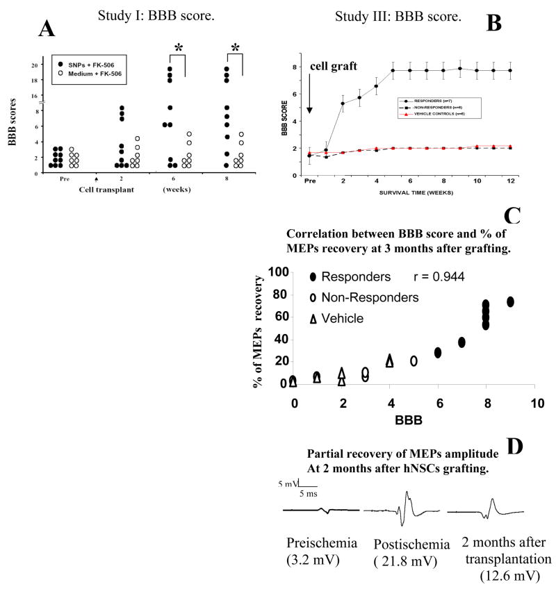
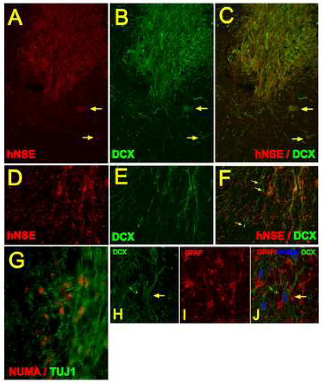
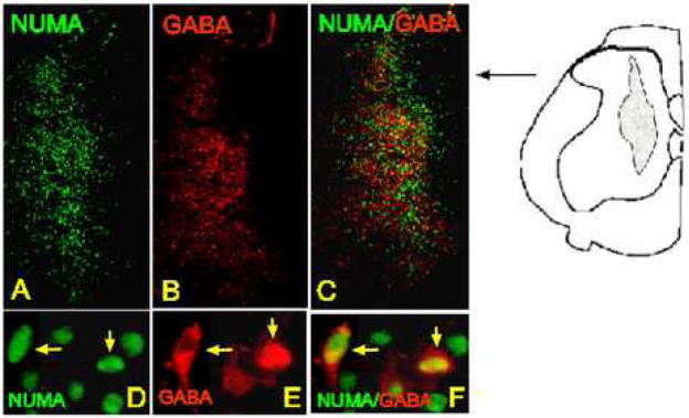
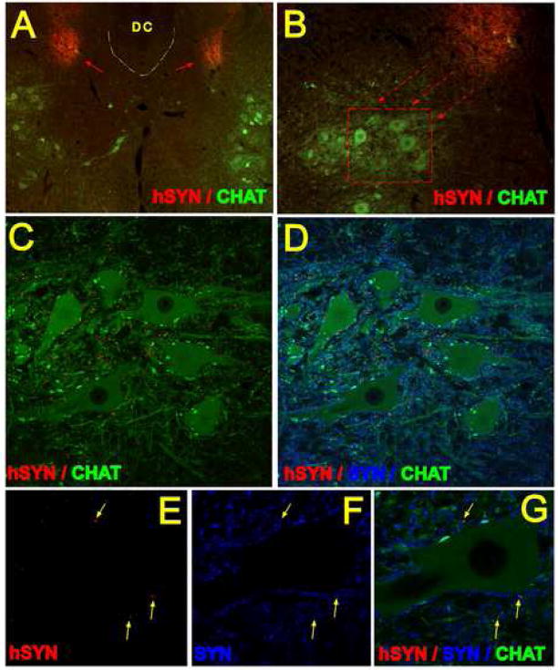
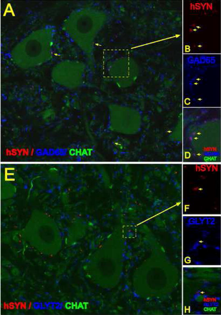
Transient spinal cord ischemia in humans can lead to the development of permanent paraplegia with prominent spasticity and rigidity. Histopathological analyses of spinal cords in animals with ischemic spastic paraplegia show a selective loss of small inhibitory interneurons in previously ischemic segments but with a continuing presence of ventral alpha-motoneurons and descending cortico-spinal and rubro-spinal projections. The aim of the present study was to examine the effect of human spinal stem cells (hSSCs) implanted spinally in rats with fully developed ischemic paraplegia on the recovery of motor function and corresponding changes in motor evoked potentials. In addition the optimal time frame for cell grafting after ischemia and the optimal dosing of grafted cells were also studied. Spinal cord ischemia was induced for 10 min using aortic occlusion and systemic hypotension. In the functional recovery study, hSSCs (10,000-30,000 cells/0.5 mul/injection) were grafted into spinal central gray matter of L2-L5 segments at 21 days after ischemia. Animals were immunosuppressed with Prograf (1 mg/kg or 3 mg/kg) for the duration of the study. After cell grafting the recovery of motor function was assessed periodically using the Basso, Beattie and Bresnahan (BBB) scoring system and correlated with the recovery of motor evoked potentials. At predetermined times after grafting (2-12 weeks), animals were perfusion-fixed and the survival, and maturation of implanted cells were analyzed using antibodies recognizing human-specific antigens: nuclear protein (hNUMA), neural cell adhesion molecule (hMOC), neuron-specific enolase (hNSE) and synapthophysin (hSYN) as well as the non-human specific antibodies TUJ1, GFAP, GABA, GAD65 and GLYT2. After cell grafting a time-dependent improvement in motor function and suppression of spasticity and rigidity was seen and this improvement correlated with the recovery of motor evoked potentials. Immunohistochemical analysis of grafted lumbar segments at 8 and 12 weeks after grafting revealed intense hNSE immunoreactivity, an extensive axo-dendritic outgrowth as well as rostrocaudal and dorsoventral migration of implanted hNUMA-positive cells. An intense hSYN immunoreactivity was identified within the grafts and in the vicinity of persisting alpha-motoneurons. On average, 64% of hSYN terminals were GAD65 immunoreactive which corresponded to GABA immunoreactivity identified in 40-45% of hNUMA-positive grafted cells. The most robust survival of grafted cells was seen when cells were grafted 21 days after ischemia. As defined by cell survival and laminar distribution, the optimal dose of injected cells was 10,000-30,000 cells per injection. These data indicate that spinal grafting of hSSCs can represent an effective therapy for patients with spinal ischemic paraplegia.


Figures

Figure 1 A, B, C
Figure 1 A, B, C
hSSCs co-cultured on a rat astrocyte monolayer for 6 weeks and stained with human-specific NSE antibody (A) and GABA antibody (B). Extensive axodendritic sprouting and GABA immunoreactivity in terminally differentiated neurons was noted.
Figure 2 A – N
Figure 2 A – N
A, B: Transverse lumbar spinal cord sections taken at 7 weeks after grafting from an animal injected with 30 000 cells/injection. Staining with human specific nuclear antibody (NUMA) confirmed the presence of grafted cells distributed between the superficial dorsal horn layers and lamina VIII. C, D: Staining with human specific MOC and NSE antibody revealed intense immunoreactivity within the individual grafts. E, F, G: Sagittal lumbar spinal cord sections stained with NUMA and MOC antibody. The core of the individual injection sites could clearly be recognized (E, F, G: asterisks). Numerous NUMA- and MOC-positive cells which migrated for distances greater then 500μm from the borders of the grafts can also be identified (white arrows). H, I, J, K: Confocal analysis of NUMA and GFAP antibody-stained sections showed that a sub-population of NUMA-positive cells differentiated into astrocytes (I, J, K). These cells were typically localized at the periphery of the individual grafts. L, M, N: Systematic 3-D analysis of serial transverse spinal cord sections stained with the hMOC antibody confirmed a consistent presence of MOC-positive grafts across 3 individual injections sites (DH-dorsal horn; VH-ventral horn).
Figure 3 A – D
Figure 3 A – D
Recovery of motor function and motor evoked potentials in animals grafted with hSSCs. A: Statistical analysis of the BBB score between groups A3 and A4 (Study I) revealed a significant improvement at 6 and 8 weeks (P<0.05). B: In Study III 7 of 13 animals grafted with hSSCs showed an improvement in the movement of all 3 joints of the lower extremities and relief of muscle spasticity and rigidity. Six animals showed no improvement. Correlative assessment with the degree of BBB recovery and the degree of MEPs normalization showed a significant correlation (C). D: MEPs recording in an individual animal at baseline, after ischemia and at 2 months after hSSCs grafting. Increased MEPs amplitudes were found after ischemia, followed by a partial normalization at 2 months after grafting.
Figure 4 A – J
Figure 4 A – J
Transverse spinal cord sections taken at 3 months after grafting and double stained with human specific NSE antibody (A-red) and DCX antibody (B-green). Intense NSE and DCX staining was seen within the graft. In a number of grafted terminally differentiated neurons the colocalization of both proteins was identified (A, B, C- yellow arrows). Numerous processes were found extending from the graft which were DCX immunoreactive but NSE negative (D, E, F; yellow arrows). Double staining with NUMA and TUJ1 antibody revealed that virtually all NUMA immunoreactive cells were also TUJ1-positive (G). H, I, J- Projected confocal images taken from sections triple stained with DCX, NUMA and non-human specific GFAP antibody. Numerous host-derived GFAP-positive astrocytes which were NUMA-negative were identified within the graft. DCX and NUMA-labeled neurons with extensive axo-dendritic sprouting can also be seen (H, I, J-yellow arrow).
Figure 5 A – F
Figure 5 A – F
Transverse spinal cord section taken after 7 weeks of survival and stained with NUMA and GABA antibody. A subpopulation of NUMA-positive cells showed colocalization with GABA immunoreactivity (D, E, F: yellow arrows).
Figure 6 A – G
Figure 6 A – G
A, B, C - Fluorescent microscopy images (A, B) and projected confocal images (C, D) of transverse spinal cord sections taken at 3 months after grafting and stained with human specific synaptophysin antibody (red), CHAT antibody (green) and synaptophysin antibody which cross-reacts with both human and rat synaptohysin (SYN; blue). Intense hSYN immunoreactivity was found within the two bilateral grafts (A; red arrows). Numerous hSYN immunoreactive terminals were localized in the base of the dorsal horn and extending into ventral α-motoneuron pools (B, C; red). E, F, G - single optical images (0.3μm thick) demonstrating colocalization of hSYN and SYN immunoreactivity in the vicinity of persisting CHAT+ α-motoneuron (yellow arrows).
Figure 7 A – H
Figure 7 A – H
A, B, C, D- single confocal optical images taken from section at 3 months after grafting and stained with hSYN (red), CHAT (green) and GAD65 (blue) antibodies. The majority of hSYN terminals showed colocalization with GAD65 (B, C, D-yellow arrows). E, F, G, H - single confocal optical images taken from sections at 3 months after grafting and stained with hSYN (red), CHAT (green) and GLYT2 (blue) antibodies. Only occasional colocalization of hSYN and GLYT2 antibody was noted (F, G, H - yellow arrow).

References

    1. Acsadi G, Anguelov RA, Yang H, Toth G, Thomas R, Jani A, Wang Y, Ianakova E, Mohammad S, Lewis RA, Shy ME. Increased survival and function of SOD1 mice after glial cell-derived neurotrophic factor gene therapy. Hum Gene Ther. 2002;13:1047–1059. - PubMed
    1. Akins PT, Atkinson RP. Glutamate AMPA receptor antagonist treatment for ischaemic stroke. Curr Med Res Opin. 2002;18(Suppl 2):s9–13. - PubMed
    1. Anderson DK, Howland DR, Reier PJ. Fetal neural grafts and repair of the injured spinal cord. Brain Pathol. 1995;5:451–457. - PubMed
    1. Basso DM, Beattie MS, Bresnahan JC, Anderson DK, Faden AI, Gruner JA, Holford TR, Hsu CY, Noble LJ, Nockels R, Perot PL, Salzman SK, Young W. MASCIS evaluation of open field locomotor scores: effects of experience and teamwork on reliability. Multicenter Animal Spinal Cord Injury Study. Journal of Neurotrauma. 1996;13:343–359. - PubMed
    1. Bendel O, Bueters T, von Euler M, Ove Ogren S, Sandin J, von Euler G. Reappearance of hippocampal CA1 neurons after ischemia is associated with recovery of learning and memory. J Cereb Blood Flow Metab 2005 - PubMed

Publication types

MeSH terms