

Injections of NGF into neonatal frontal cortex decrease social interaction as adults: a rat model of schizophrenia
- PMID: 17525084
- PMCID: PMC2632378
- DOI: 10.1093/schbul/sbm039
Injections of NGF into neonatal frontal cortex decrease social interaction as adults: a rat model of schizophrenia
Abstract
Background: Injection of nerve growth factor (NGF) into the developing frontal cortex (FC) has been shown to produce adult-onset subcortical dopaminergic hyperactivity, impaired prepulse inhibition of the acoustic startle response, and several neuropathological features of schizophrenia. The present study was to determine whether such lesions would lead to impaired social interaction, a prominent negative feature of schizophrenia.
Methods: Rat pups received daily injections of human recombinant NGF into the developing FC on postnatal days 1 and 2 to partially lesion subplate neurons. Lesioned rats were tested in similar-treatment pairings lasting 23.5 hours using the EthoVision behavioral monitoring system at 6 and 14 weeks of age. Brains were then perfusion fixed for histological analysis.
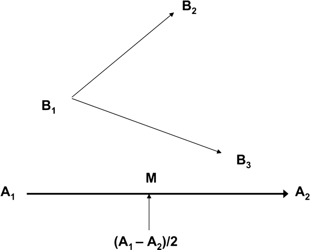
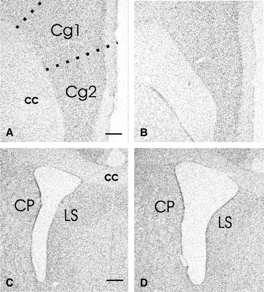
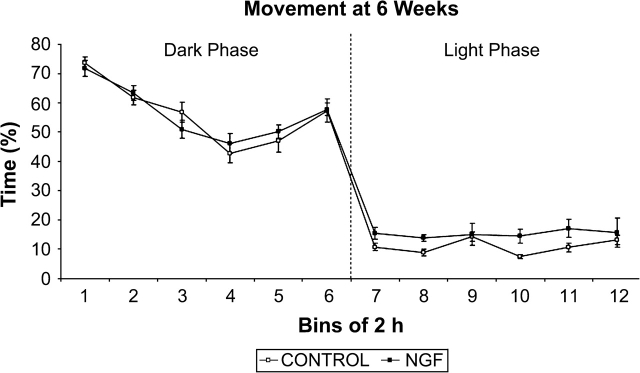
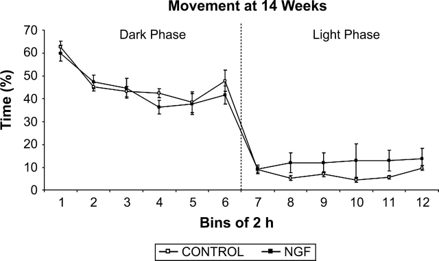
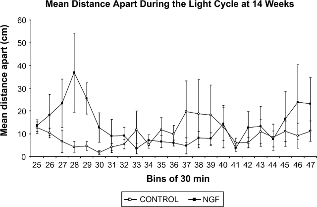
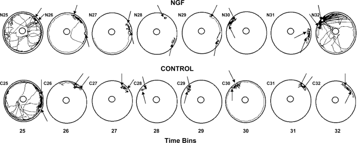
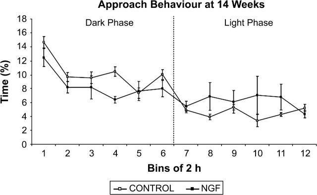
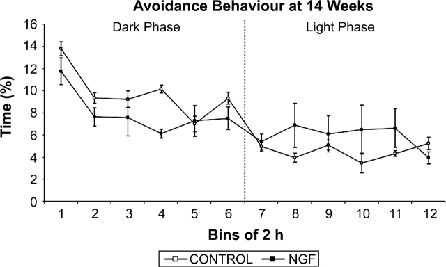
Results: Lesioned rats showed significantly increased movement, relative to controls, during the light phase at 6 weeks of age. At 14 weeks, they maintained a significantly greater mean distance apart from one another, and engaged in significantly less approach and avoidance behavior during the dark phase, relative to controls. Histological changes were consistent with those described previously in this animal model.
Conclusion: Results indicate that injections of NGF into the developing FC of neonatal rats result in reduced social interaction, which is consistent with behaviors observed in human schizophrenia patients.
Figures

Similar articles
-
Infusions of Nerve Growth Factor Into the Developing Frontal Cortex Leads to Deficits in Behavioral Flexibility and Increased Perseverance.Schizophr Bull. 2018 Aug 20;44(5):1081-1090. doi: 10.1093/schbul/sbx159. Schizophr Bull. 2018. PMID: 29165654 Free PMC article.
-
Altered neurotrophin receptor function in the developing prefrontal cortex leads to adult-onset dopaminergic hyperresponsivity and impaired prepulse inhibition of acoustic startle.Biol Psychiatry. 2004 Apr 15;55(8):797-803. doi: 10.1016/j.biopsych.2003.12.015. Biol Psychiatry. 2004. PMID: 15050860
-
Alterations to prepulse inhibition magnitude and latency in adult rats following neonatal treatment with domoic acid and social isolation rearing.Behav Brain Res. 2016 Feb 1;298(Pt B):310-7. doi: 10.1016/j.bbr.2015.11.009. Epub 2015 Nov 15. Behav Brain Res. 2016. PMID: 26590368
-
Evaluation of the antipsychotic effect of bi-acetylated l-stepholidine (l-SPD-A), a novel dopamine and serotonin receptor dual ligand.Schizophr Res. 2009 Nov;115(1):41-9. doi: 10.1016/j.schres.2009.08.002. Epub 2009 Sep 9. Schizophr Res. 2009. PMID: 19744833
-
The role of the hippocampo-prefrontal cortex system in phencyclidine-induced psychosis: a model for schizophrenia.J Physiol Paris. 2013 Dec;107(6):434-40. doi: 10.1016/j.jphysparis.2013.06.002. Epub 2013 Jun 17. J Physiol Paris. 2013. PMID: 23792022 Review.
Cited by
-
Cross-species analyses of the cortical GABAergic and subplate neural populations.Front Neuroanat. 2009 Oct 6;3:20. doi: 10.3389/neuro.05.020.2009. eCollection 2009. Front Neuroanat. 2009. PMID: 19936319 Free PMC article.
-
SELENBP1 overexpression in the prefrontal cortex underlies negative symptoms of schizophrenia.Proc Natl Acad Sci U S A. 2022 Dec 20;119(51):e2203711119. doi: 10.1073/pnas.2203711119. Epub 2022 Dec 13. Proc Natl Acad Sci U S A. 2022. PMID: 36512497 Free PMC article.
-
Contribution of nonprimate animal models in understanding the etiology of schizophrenia.J Psychiatry Neurosci. 2011 Jul;36(4):E5-29. doi: 10.1503/jpn.100054. J Psychiatry Neurosci. 2011. PMID: 21247514 Free PMC article. Review.
-
Dysregulation of mTOR signaling mediates common neurite and migration defects in both idiopathic and 16p11.2 deletion autism neural precursor cells.Elife. 2024 Mar 25;13:e82809. doi: 10.7554/eLife.82809. Elife. 2024. PMID: 38525876 Free PMC article.
-
[Are psychic disorders specifically human?].Nervenarzt. 2009 Mar;80(3):252-62. doi: 10.1007/s00115-008-2591-2. Nervenarzt. 2009. PMID: 19011825 Review. German.
References
-
- American Psychiatric Association. Diagnostic and Statistical Manual of Mental. 4th ed. Washington, DC: American Psychiatric Press; 2000.
-
- Weinberger DR. Implications of normal brain development for the pathogenesis of schizophrenia. Arch Gen Psychiatry. 1987;43:114–124. - PubMed
-
- Bediou B, Franck N, Saoud M, et al. Effects of emotion and identity on facial affect processing in schizophrenia. Psychiatry Res. 2005;133:149–157. - PubMed
-
- Bellack AS, Morrison RL, Wixted JT, Mueser KT. An analysis of social competence in schizophrenia. Br J Psychiatry. 1990;156:809–818. - PubMed
Publication types
MeSH terms
Substances
LinkOut - more resources
Full Text Sources
Other Literature Sources
Medical
