. 2007 Aug 1;582(Pt 3):1205-17.
doi: 10.1113/jphysiol.2007.134809. Epub 2007 May 24.

Membrane cholesterol modulates Kv1.5 potassium channel distribution and function in rat cardiomyocytes

Affiliations

Membrane cholesterol modulates Kv1.5 potassium channel distribution and function in rat cardiomyocytes

Joëlle Abi-Char et al. J Physiol. .

Abstract

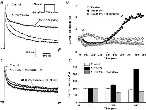
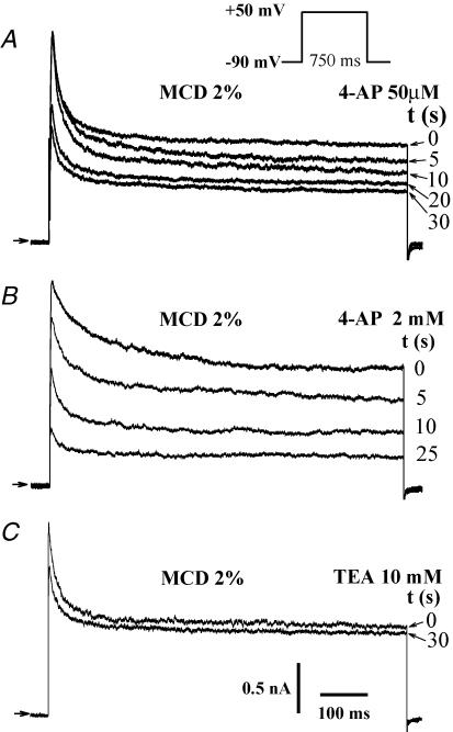
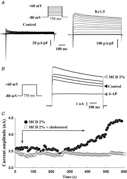
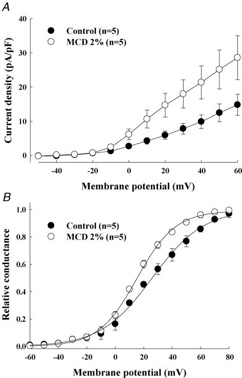
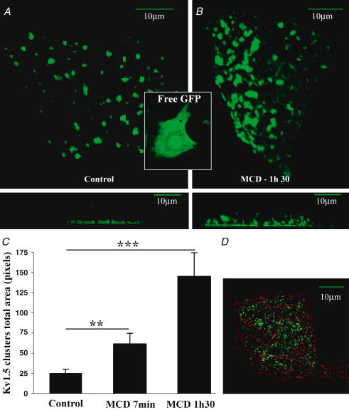
Membrane lipid composition is a major determinant of cell excitability. In this study, we assessed the role of membrane cholesterol composition in the distribution and function of Kv1.5-based channels in rat cardiac membranes. In isolated rat atrial myocytes, the application of methyl-beta-cyclodextrin (MCD), an agent that depletes membrane cholesterol, caused a delayed increase in the Kv1.5-based sustained component, I(kur), which reached steady state in approximately 7 min. This effect was prevented by preloading the MCD with cholesterol. MCD-increased current was inhibited by low 4-aminopyridine concentration. Neonatal rat cardiomyocytes transfected with Green Fluorescent Protein (GFP)-tagged Kv1.5 channels showed a large ultrarapid delayed-rectifier current (I(Kur)), which was also stimulated by MCD. In atrial cryosections, Kv1.5 channels were mainly located at the intercalated disc, whereas caveolin-3 predominated at the cell periphery. A small portion of Kv1.5 floated in the low-density fractions of step sucrose-gradient preparations. In live neonatal cardiomyocytes, GFP-tagged Kv1.5 channels were predominantly organized in clusters at the basal plasma membrane. MCD caused reorganization of Kv1.5 subunits into larger clusters that redistributed throughout the plasma membrane. The MCD effect on clusters was sizable 7 min after its application. We conclude that Kv1.5 subunits are concentrated in cholesterol-enriched membrane microdomains distinct from caveolae, and that redistribution of Kv1.5 subunits by depletion of membrane cholesterol increases their current-carrying capacity.


Figures

Figure 1
Figure 1. Effects of cholesterol depletion on outward currents in native adult atrial myocytes
A, direct and long period effects of MCD application on the outward current elicited as indicated by the command waveform at 0.2 Hz, shown in the inset. B, effects of MCD–cholesterol application. C, time course changes in Ikur amplitude in control conditions, upon application of MCD and MCD–cholesterol complexes. D, bar graphs summarizing the effects of MCD and MCD–cholesterol complex, measured after 700 s application.
Figure 2
Figure 2. Pharmacological properties of the MCD-activated current in native adult atrial myocytes
Effects of 50 μm (A) and 2 mm 4-AP (B), and 10 mm TEA (C), on the 650 s MCD-stimulated outward current elicited as indicated by the command waveform at 0.2 Hz.
Figure 3
Figure 3. MCD effects on electrophysiological parameters of the outward K+ current of native adult atrial myocytes
Current traces elicited during incremental depolarizing test pulses in control (A) conditions and after MCD application (B). Cell capacitance: 75 pF. C, current density–voltage relationships of Ikur recorded in control conditions (•) and following MCD (^) application. D, voltage dependence of current activation in control (•) and MCD (^) conditions. In C and D, each point is the average of 7 cells.
Figure 4
Figure 4. MCD enhances Kv1.5-encoded currents in neonatal cardiomyocytes
A, traces of currents recorded during incremental 10 mV step depolarizations in control and in Kv1.5-transfected cardiomyocytes. B, effects of MCD on the outward current elicited by a test pulse from −80 to +60 mV in control, and after 350, 500 and 600 s MCD applications. Effect of 500 μm 4-AP on 650 s MCD-stimulated current is also shown. C, time course of the MCD and MCD–cholesterol complex effects on the outward current.
Figure 5
Figure 5. Effect of cholesterol depletion on outward current parameters resulting from Kv1.5 subunit overexpression in neonatal cardiomyocytes
Current density–voltage relationships (A) and voltage dependence (B) of IKur activation under control conditions (•) and following 7 min MCD application (^). In A and B, each point represents average data from 5 cells.
Figure 6
Figure 6. Kv1.5 do not co-localized with caveolin-3 in adult atrial tissue
Immunolocalization of Kv1.5 subunits (A) and caveolin-3 (B) in cryosections of atrial myocardium. Double immunostaining of connexin-43 (FITC, C) and caveolin-3 (Texas Red, D) in cryosections of atrial myocardium. E, merged image of the same area of the section in C and D, showing the lack of overlap between connexin-43 and caveolin-3 stainings.
Figure 7
Figure 7. Some Kv1.5 channel subunits are localized in lipid raft fractions in adult rat atrial myocytes
Western blot analysis of the distribution of Kv1.5 subunits, connexin-43 and caveolin-3 on step sucrose gradient of proteins from atrial myocardium.
Figure 8
Figure 8. Surface expression of Kv1.5 subunits in neonatal cardiomyocytes
A, in live cardiomyocytes, transfected GFP-tagged Kv1.5 subunits are clustered at the membrane surface adjacent to the bottom of laminin-coated glass support, as shown in the projection of Z sections in the lower panel. In contrast, GFP alone was homogeneously distributed in cardiomyocytes (inset). B, after the application of 2% MCD, clusters increased in size and were redistributed throughout the plasma membrane. C, bar graphs summarizing changes in cluster size upon MCD exposures; data are from 21 cardiomyocytes in control, and following incubation with 2% MCD for 7 min and 1 h 30 min. **P < 0.01, ***P < 0.001. D, double immunostaining of fixed cardiomyocytes using sarcomeric α-actinin and anti-Kv1.5 antibodies showing that Kv1.5-GFP-transfected cells are cardiomyocytes. A, B and D: scale bars represent 10 μm.

References

    1. Barbuti A, Gravante B, Riolfo M, Milanesi R, Terragni B, DiFrancesco D. Localization of pacemaker channels in lipid rafts regulates channel kinetics. Circ Res. 2004;94:1325–1331. - PubMed
    1. Boixel C, Gonzalez W, Louedec L, Hatem SN. Mechanisms of L-type Ca2+ current downregulation in rat atrial myocytes during heart failure. Circ Res. 2001;89:607–613. - PubMed
    1. Brainard AM, Miller AJ, Martens JR, England SK. Maxi-K channels localize to caveolae in human myometrium: a role for an actin-channel-caveolin complex in the regulation of myometrial smooth muscle K+ current. Am J Physiol Cell Physiol. 2005;289:C49–C57. - PubMed
    1. Christian AE, Haynes MP, Phillips MC, Rothblat GH. Use of cyclodextrins for manipulating cellular cholesterol content. J Lipid Res. 1997;38:2264–2272. - PubMed
    1. Czub J, Baginski M. Comparative molecular dynamics study of lipid membranes containing cholesterol and ergosterol. Biophys J. 2006;90:2368–2382. - PMC - PubMed

Publication types