. 2007 Jun;48(6):2576-88.
doi: 10.1167/iovs.06-0662.

Horizontal rectus muscle anatomy in naturally and artificially strabismic monkeys

Affiliations

Horizontal rectus muscle anatomy in naturally and artificially strabismic monkeys

Anita Narasimhan et al. Invest Ophthalmol Vis Sci. 2007 Jun.

Abstract

Purpose: Structural abnormalities of extraocular muscles (EOMs) or their pulleys are associated with some forms of human strabismus. This experiment was conducted to investigate whether such abnormalities are associated with artificial or naturally occurring strabismus in monkeys.

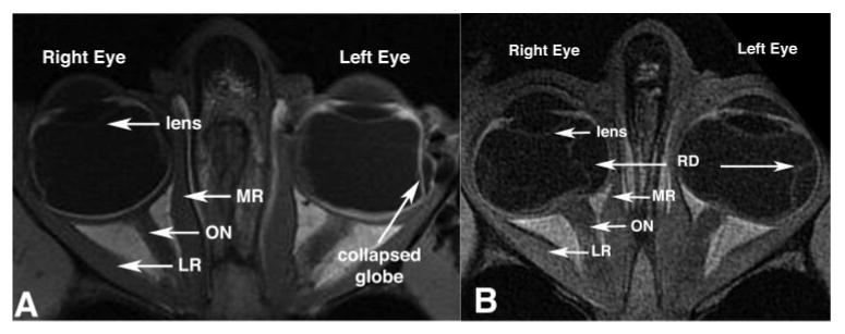
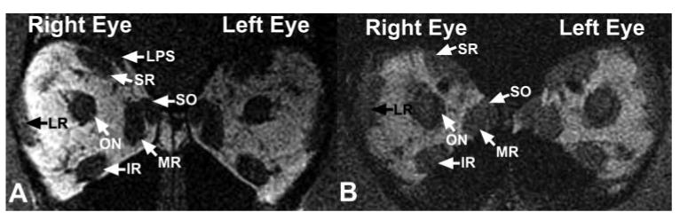
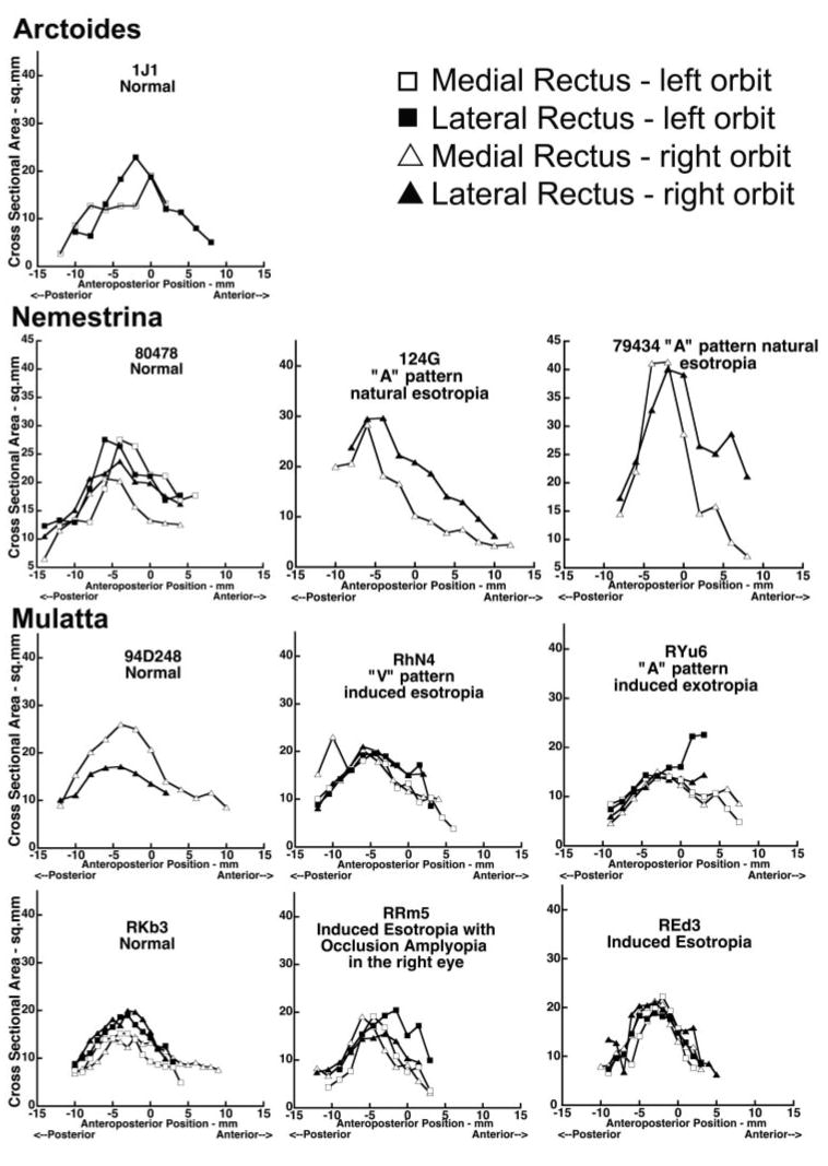
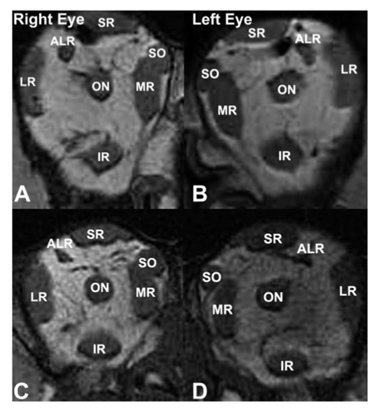
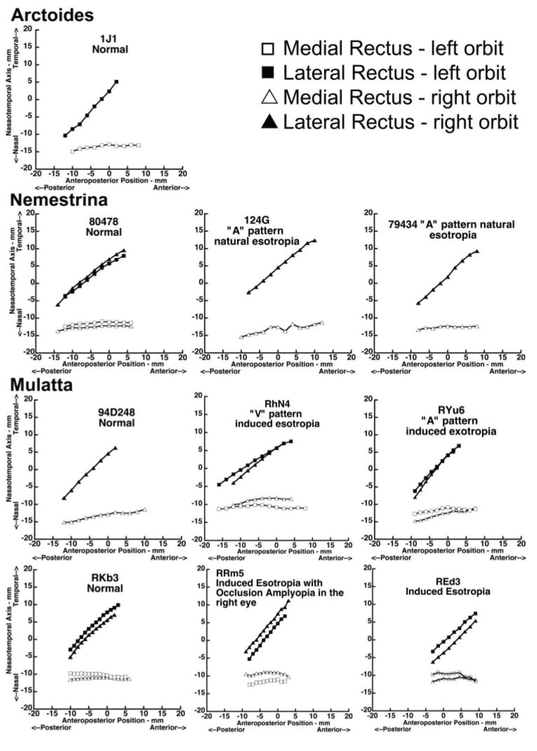
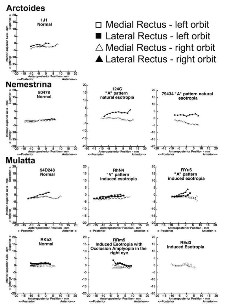
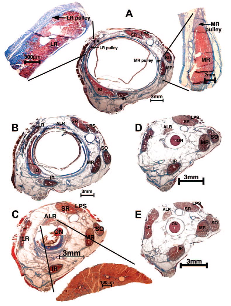
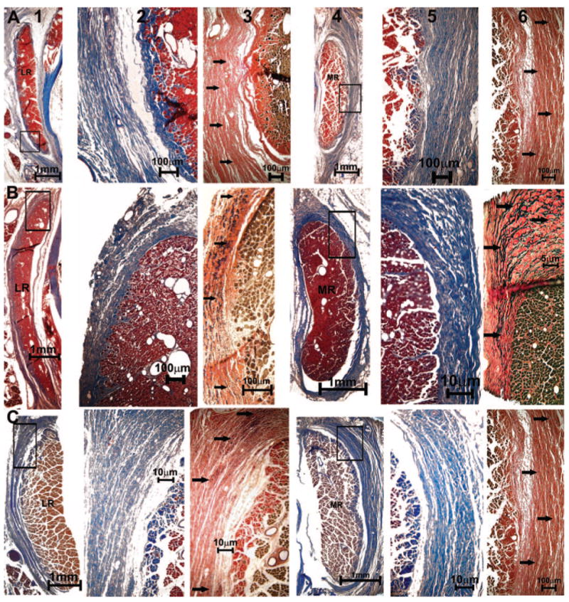
Methods: Binocular alignment and grating visual acuities were determined in 10 monkeys representing various species using search coil recording and direct observations. Four animals were orthotropic, two had naturally occurring "A"-pattern esotropia, two had concomitant and one had "V"-pattern esotropia artificially induced by alternating or unilateral occlusion in infancy, and one had "A"-pattern exotropia artificially induced by prism wear. After euthanasia, 16 orbits were examined by high-resolution magnetic resonance imaging (MRI) in the quasi-coronal plane. Paths and sizes of horizontal rectus EOMs were analyzed quantitatively in a standardized coordinate system. Whole orbits were then serially sectioned en bloc in the quasi-coronal plane, stained for connective tissue, and compared with MRI. Nerve and EOM features were analyzed quantitatively.

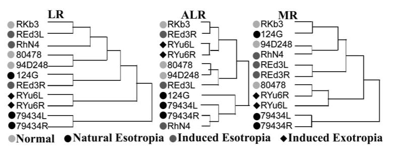
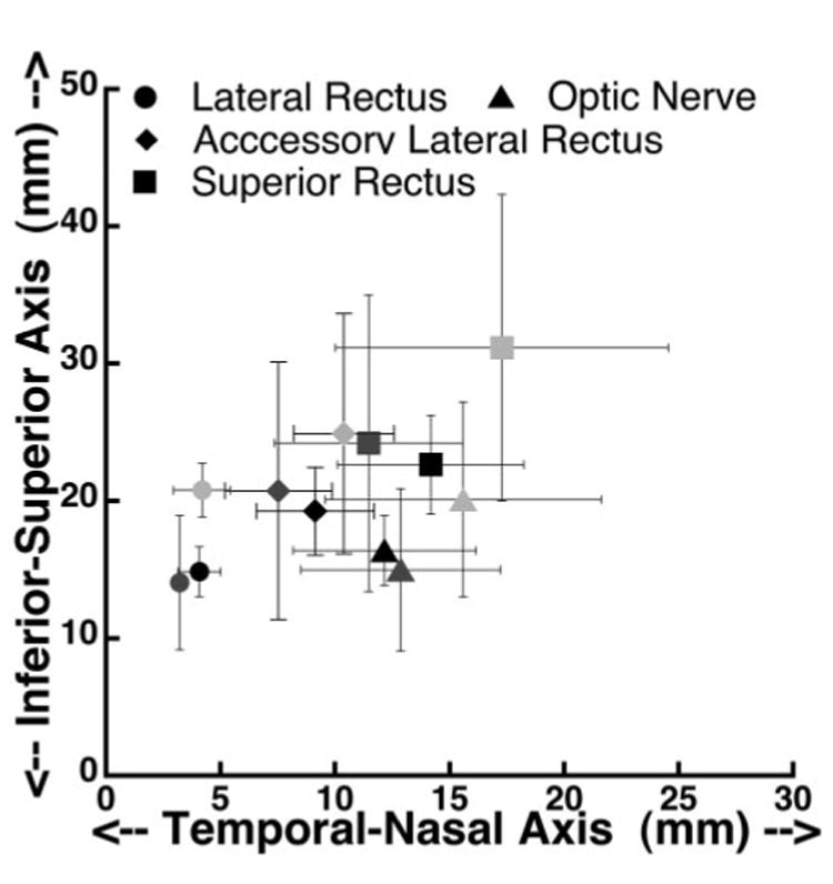
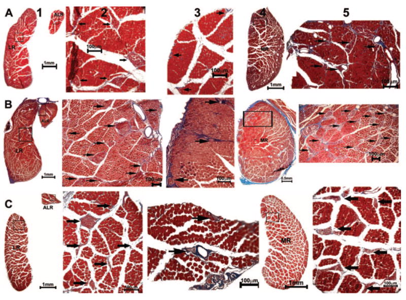
Results: Quantitative analysis of MRI revealed no significant differences in horizontal rectus EOM sizes or paths among orthotropic or naturally or artificially strabismic monkeys. Histologic examination demonstrated no differences in EOM size, structure, or innervation among the three groups, and no differences in connective tissues in the pulley system. The accessory lateral rectus (ALR) EOM was present in all specimens, but was small, inconsistently located, and sparsely innervated. Characteristics of the ALR did not correlate with strabismus.

Conclusions: Major structural abnormalities of horizontal rectus EOMs and associated pulleys are unrelated to natural or artificial horizontal strabismus in the monkeys studied. The ALR is unlikely to contribute to horizontal strabismus in primates. However, these findings do not exclude a possible role of pulley abnormalities in disorders such as cyclovertical strabismus.


Figures

Figure 1
Figure 1
Axial MRIs showed no systematic differences between the orbits of orthotropic and strabismic monkeys. Left: orthotropic M. mulatta 94D248. Resolution, 273 μm in a 3-mm-thick plane. Right: artificially induced right esotropic M. mulatta RRm5, which had undergone monocular occlusion of the right eye. Resolution, 234 μm in 2-mm-thick plane. RD, retinal detachment.
Figure 2
Figure 2
Quasicoronal MRIs taken 9 mm posterior to the globe- optic nerve junction demonstrated no systematic differences between orbits of normal and strabismic monkeys. Left: orthotropic Macaca nemestrina 80478. Resolution, 312 μm in 2-mm-thick image planes. Right: naturally esotropic M. nemestrina 124G. Resolution, 390 μm in 3-mm-thick image planes. IR, inferior rectus muscle; LPS, levator palpebrae superioris muscle.
Figure 3
Figure 3
Cross-sectional areas of the MR and LR in normal and strabismic monkeys plotted against anteroposterior distance along the muscles, relative to 0 at the globe- optic nerve junction.
Figure 4
Figure 4
Hierarchical clustering analysis based on cross-sectional areas of the MR, LR, and ALR in normal and strabismic monkeys. Clustering did not occur according to binocular alignment or laterality, species, age, or gender.
Figure 5
Figure 5
Quasicoronal MRI of the right and left orbits of monkeys 1.5 to 2 mm posterior to the globe- optic nerve junction. (A, B) Orthotropic M. mulatta 94D248. Resolution, 234 μm in 2-mm-thick image planes. (C, D) Induced exotropic M. mulatta RYu6. Resolution, 234 μm in 1.5-mm-thick image planes. IR, inferior rectus muscle.
Figure 6
Figure 6
Horizontal coordinates of horizontal rectus EOM paths along the orbit for both normal and strabismic monkeys, 0 at the globe- optic nerve junction.
Figure 7
Figure 7
Vertical coordinates of horizontal rectus EOM paths for the normal and strabismic monkeys. Anteroposterior positions are relative to 0, at the globe- optic nerve junction.
Figure 8
Figure 8
Location of accessory lateral rectus muscle at the anteroposterior level of the globe and optic nerve junction, in comparison to the optic ON and the LR and SR muscles. Light gray symbols: normal monkeys; black symbols: esotropic monkeys; dark gray symbols: exotropic monkey.
Figure 9
Figure 9
Histologic sections in the quasicoronal plane from right orbit of naturally esotropic monkey 124G stained with MT. (A) In the pulley region 4 mm anterior to globe- optic nerve junction. The LR and MR muscles are magnified in adjacent insets. (B) Two millimeters anterior to the globe- optic nerve junction. This region is immediately posterior to the rectus pulleys. (C) At the globe- optic nerve junction, well posterior to the pulleys. Inset: magnified view of the ALR. (D) Two millimeters and (E) 4 mm posterior to the globe- optic nerve junction. LPS, levator palpebrae superioris muscle; IR, inferior rectus muscle.
Figure 10
Figure 10
Quasicoronal histologic sections of monkey orbits stained with MT and EVG stains. (A) Normal (94D248 right orbit), (B) naturally esotropic (124G left orbit), and (C) artificially esotropic (RHn4 left orbit) monkeys. Columns from left to right: lateral rectus (LR) EOM (1), magnified inset of pulley in MT stain (2), and elastin fibrils (arrows) in EVG stain (3), MR EOM (4), magnified inset of collagen fibers in MT stain (5), and elastin fibrils (arrows) in EVG stain (6).
Figure 11
Figure 11
Quasicoronal histologic sections from (A) normal (94D248 left orbit, 19.16 mm from the cornea), (B) naturally esotropic (124G right orbit, 23.61 mm from cornea), and (C) artificially esotropic (RhN4 left orbit, 20.97 mm from cornea) monkeys, stained with MT. Columns from left to right: LR and ALR EOMs (1) with intramuscular nerve arborization (2, 3); MR (4) and its innervation (5). Arrows: nerves.

References

    1. Miller JM. Computer model of binocular alignment. In: Semmlow JL, Welkowitz W, editors. Sixth Annual Conference, IEEE Engineering in Medicine and Biology Society. New York: IEEE; 1984.
    1. Miller JM, Pavlovski DS, Shaemeva I. Orbit 1.8 Gaze Mechanics Simulation. San Francisco: Eidactics; 1999.
    1. Robinson DA. A quantitative analysis of extraocular muscle cooperation and squint. Invest Ophthalmol Vis Sci. 1975;14:801–825. - PubMed
    1. Miller JM, Robins D. Extraocular muscle sideslip and orbital geometry in monkeys. Vision Res. 1987;27:381–392. - PubMed
    1. Simonsz HJ, Harting F, de Waal BJ, Verbeeten BWJM. Sideways displacement and curved path of recti eye muscles. Arch Ophthalmol. 1985;103:124–128. - PubMed

Publication types

LinkOut - more resources