. 2007 Jun;48(6):2868-76.
doi: 10.1167/iovs.06-0035.

Transport of truncated rhodopsin and its effects on rod function and degeneration

Affiliations

Transport of truncated rhodopsin and its effects on rod function and degeneration

Edwin S Lee et al. Invest Ophthalmol Vis Sci. 2007 Jun.

Abstract

Purpose: Most transgenic animal models of retinal degeneration caused by rhodopsin mutations express the rhodopsin transgene on a wild-type (WT) genetic background. Previous studies have demonstrated that one mechanism of retinal degeneration is rhodopsin overexpression. To study the effect of C-terminal truncation of rhodopsin without the confounding factors of overexpression, several lines of transgenic mice were generated that expressed a C-terminal rhodopsin mutation on rhodopsin-knockout backgrounds.

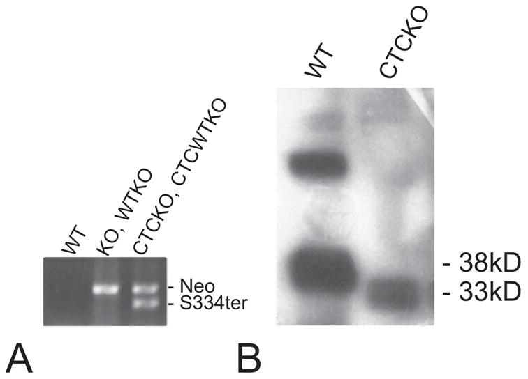
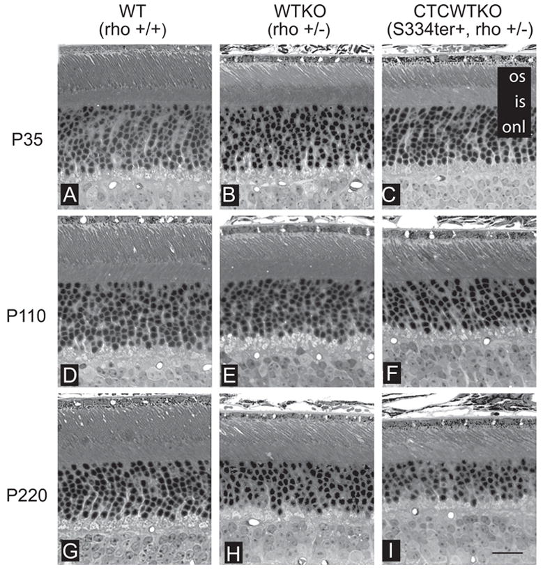
Methods: Two lines of transgenic mice, expressing different levels of C-terminal truncated rhodopsin (S334ter) were mated with heterozygous rhodopsin-knockout (rho+/-) mice to express S334ter rhodopsin on a background with reduced endogenous rhodopsin expression. S334ter mice were mated to homozygous knockout (rho-/-) mice to examine the effect of S334ter rhodopsin on a null rhodopsin background. S334ter rhodopsin expression was estimated by Western blot. Retinal function was assessed by ERG and retinal degeneration by histopathology and morphometry. C-terminal rhodopsin sorting and trafficking was examined by fluorescence immunocytochemistry with detection by electron microscope.

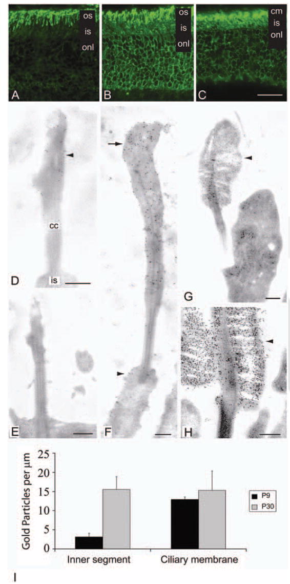
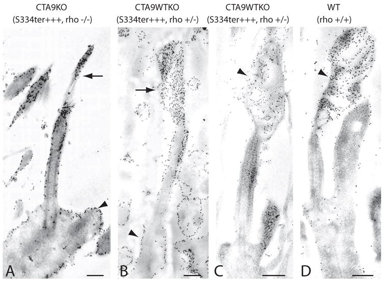
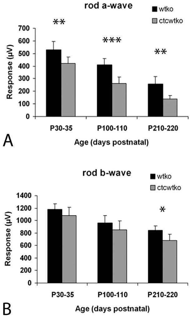
Results: Expression of S334ter truncated rhodopsin at low levels in the presence of decreased total rhodopsin in rods (S334ter, rho+/-) increased the rate of rod cell death in comparison to rho+/- littermates. In addition, S334ter rhodopsin prolonged the recovery time of the rod ERG to a light flash and diminished the a-wave amplitudes in comparison to their (rho+/-) littermates. Photoreceptors of S334ter mice on a homozygous rhodopsin-knockout background (S334ter+, rho-/-) had a fraction of mutant rhodopsin localized to the ciliary membranes.

Conclusions: Expression of S334ter rhodopsin without overexpression of total opsin in the rod photoreceptor decreased rod cell contribution to the ERG and compromised rod cell survival in adult mice. The increased cell death may be a consequence of C-terminal truncated rhodopsin mislocalization in membranes of the inner segment. Another possible pathologic mechanism is prolonged activation of phototransduction from the presence of mutant rhodopsin in the outer segment lacking the normal C-terminal binding sites for shutoff by arrestin and phosphorylation. These results suggest that rhodopsin lacking a C-terminal trafficking signal can be transported to the rod outer segment without cotransporting with full-length rhodopsin.


Figures

Figure 1
Figure 1
Western blot and transgene PCR analysis of WT and mutant mice. (A) PCR analysis of genomic DNA with primers specific for the Neomycin (Neo) KO cassette and the S334ter transgene confirmed their presence or absence. KO (rho−/−) and WTKO (rho+/−) mice are positive for the neomycin cassette. CTCKO (S334ter+, rho+/−) and CTCWTKO (S334ter+, rho−/−) animals are positive for both Neo and S334ter PCR products. WT (rho+/+) animals are negative for either product. (B) Anti-rhodopsin antibody Rho4D2 was used to probe immunoblots prepared from P20 WT and CTCKO (S334ter+, rho−/−) retinas. S334ter rhodopsin migrated as a smaller fragment due to the removal of 15 amino acids from its C terminus.
Figure 2
Figure 2
Rhodopsin distribution in WT and mutant photoreceptors. (AC) Immunohistochemical labeling with anti-rhodopsin antibody, Rho4D2 on frozen sections of P23 WTKO, CTCWTKO, and CTCKO mice. Rho4D2 antibody recognizes the N terminus of rhodopsin and thus labels both WT and mutant forms. (A) In WTKO (rho+/−) photoreceptors, Rho4D2 predominantly immunolabeled outer segments. (B) Membranes of the outer segments, inner segments, nuclei, and synaptic terminal were labeled in the CTCWTKO (S334ter+, rho+/−) mice. (C) In CTCKO photoreceptors expressing only S334ter rhodopsin, the mutant opsin was detected in the ciliary, inner segment, perinuclear, and synaptic terminal membranes. (DH) Electron micrographs of WT and mutant photoreceptors immunolabeled with Rho4D2. (D) In P9 CTCKO photoreceptors, a low concentration of truncated rhodopsin (arrowhead) and membranes were detected in the distal connecting cilium. (E) An aged-matched rhodopsin KO photoreceptor showing no rhodopsin labeling. (F) Larger rudimentary ciliary membranes lacking organized discs were observed at P30, with higher levels of truncated S334ter rhodopsin in these distal ciliary membranes (arrow). A portion of truncated rhodopsin also mislocalized (arrowhead) to inner segment membranes. (G) Control P9 WTKO (rho+/−) photoreceptors formed small outer segments (arrowhead) heavily labeled for rhodopsin. (H) Rhodopsin localized to the rod outer segments discs (arrowhead) in P30 WTKO mice. (I) Immunogold S334ter rhodopsin labeling density in CTCKO (S334ter+, rho−/−) mice photoreceptors. Ciliary and inner segment plasma membrane densities were measured at P9 and P30. Results are expressed as the mean ± SD of three photoreceptors per age. os, outer segment; is, inner segment; onl, outer nuclear layer; cm, ciliary membrane; cc, connecting cilium. Scale bars: (A–C) 20 μm; (D–H) 250 nm.
Figure 3
Figure 3
Overexpression of truncated rhodopsin in CTA9KO and CTA9WTKO photoreceptors. Retinal sections were labeled with anti-rhodopsin antibody Rho4D2 (N-terminal) or Rho1D4 (C-terminal). (A) Only a very small concentration of membranes with truncated rhodopsin (arrow) accumulated at the connecting cilium tip in P8 CTA9KO (S334ter+++, rho−/−) photoreceptors labeled with Rho4D2. Connecting cilium and inner segment membranes (arrowhead) were densely labeled in this mouse line, which overexpressed truncated rhodopsin on a homozygous rhodopsin-knockout background. (B, C) Restoring one allele of endogenous full-length rhodopsin induced formation of small rudimentary outer segments (arrow) in a fraction of P8 CTA9WTKO photoreceptors. (B) The rhodopsin 4D2 antibody heavily labeled photoreceptor membranes (arrowhead) except for the proximal connecting cilium. (C) Anti-rhodopsin antibody, Rho1D4, recognizes residues in the C terminus of rhodopsin and specifically labeled full-length rhodopsin (arrowhead) in our animals. Endogenous, full-length rhodopsin localized to the outer segment and rarely delocalized to inner segment membranes. (D) In P8 WT photoreceptors, rhodopsin distributed predominantly to the outer segment (arrowhead), although inner segment labeling was detected in a portion of photoreceptors. Scale bars, 250 nm.
Figure 4
Figure 4
Scotopic electroretinogram recordings in WT and transgenic mice. (A, B) ERG responses after a single bright flash. (A) ERG response from CTCWTKO and WTKO animals showed a significant reduction of scotopic a-wave amplitude in CTCWTKO mice relative to WTKO mice at all ages measured. Over time, mean a-wave amplitudes decreased in both mouse lines. (B) The b-waves were significantly lower in CTCWTKO mice only at the oldest age tested (P210–P220). *P < 0.05; **P < 0.01; ***P < 0.0005 (two-tailed, unpaired t-test).
Figure 5
Figure 5
Histologic examination of WT and transgenic retinas. Light micrographs from the inferior retina were taken at different ages. (AC) P35 retinas of WT, WTKO, and CTCWTKO mice had similar ONL thicknesses (10–12 rows). Outer segments were slightly shorter in WTKO and CTCWTKO animals compared with WT controls. (DF) At P110, WTKO animals had a slightly reduced ONL thickness relative to WT animals, whereas thicknesses in CTCWTKO animals were further reduced one to two rows. Outer segments of CTCWTKO mice were occasionally shorter and slightly more disorganized than those in WTKO at this age. (GI) By P220, each genotype examined had lost one to three more rows of photoreceptors. The ONL of CTCWTKO mice was thinner, and the outer segments were generally shorter and more disorganized than in WTKO mice. Abbreviations are as in Figure 2. Scale bar, 20 μm.
Figure 6
Figure 6
Truncated rhodopsin accelerates photoreceptor degeneration in adult mice. (A) Spider graphs of ONL thickness measurements taken in 100-μm increments from the optic nerve head (ONH) to the ora serrata (ORS) in both the inferior and superior retina indicate in the P220 littermate animals that overall thickness was greatest in WT mice, followed by WTKO, and was thinnest in CTCWTKO animals. (B) Average ONL thickness plotted against age in WTKO and CTCWTKO mice. The ONL of S334ter+, rho+/− mice was significantly thinner in older animals (P110–P120 and P220–P230). Note that the x-axis is not to scale. *P < 0.005; **P < 0.001 (significant by two-tailed, unpaired t-test).

References

    1. Weleber RG, Gregory-Evans K. Retinitis pigmentosa and allied disorders. In: Ryan SJ, editor. Retina. St Louis: Mosby; 2001. pp. 362–470.
    1. The Retina Network (RetNet) Houston, TX: University of Texas-Houston Health Science Center; [Accessed June 10, 2005]. Available at http://www.sph.uth.tmc.edu/RetNet/disease/htm.
    1. Sung CH, Davenport CM, Hennessey JC, et al. Rhodopsin mutations in autosomal dominant retinitis pigmentosa. Proc Natl Acad Sci USA. 1991;88:6481–6485. - PMC - PubMed
    1. Dryja TP, Hahn LB, Cowley GS, et al. Mutation spectrum of the rhodopsin gene among patients with autosomal dominant retinitis pigmentosa. Proc Natl Acad Sci USA. 1991;88:9370–9374. - PMC - PubMed
    1. Dryja TP, McGee TL, Hahn LB, et al. Mutations within the rhodopsin gene in patients with autosomal dominant retinitis pigmentosa. N Engl J Med. 1990;323:1302–1307. - PubMed

Publication types

MeSH terms