. 2007 Jun;170(6):1917-30.
doi: 10.2353/ajpath.2007.060993.

Interferon-gamma sensitizes resistant Ewing's sarcoma cells to tumor necrosis factor apoptosis-inducing ligand-induced apoptosis by up-regulation of caspase-8 without altering chemosensitivity

Affiliations

Interferon-gamma sensitizes resistant Ewing's sarcoma cells to tumor necrosis factor apoptosis-inducing ligand-induced apoptosis by up-regulation of caspase-8 without altering chemosensitivity

Andrej Lissat et al. Am J Pathol. 2007 Jun.

Abstract

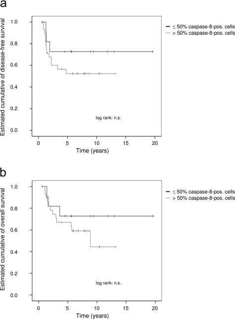
Ewing's sarcoma cells are highly susceptible to apoptosis via tumor necrosis factor apoptosis-inducing ligand (TRAIL). Resistance to TRAIL has been linked to deficient expression of caspase-8 in vitro. Here, we report on the status of caspase-8 expression in tumors from patients with Ewing's sarcoma, the effect of interferon-gamma on caspase-8 expression and apoptosis, and the role of caspase-8 for TRAIL- and chemotherapy-mediated apoptosis in Ewing's sarcoma. Using immunohistochemistry, we show that low expression of caspase-8 is seen in about 24% of tumors. Interferon-gamma induces expression of caspase-8 at concentrations achievable in humans and sensitizes cells to TRAIL. Transfection of wild type but not mutant caspase-8 into caspase-8-deficient Ewing's sarcoma cells restored sensitivity to TRAIL, indicating that up-regulation of caspase-8 is sufficient to restore TRAIL sensitivity. In contrast, no role for caspase-8 in chemotherapy-induced apoptosis was identified, because 1) transfection of caspase-8 or treatment with interferon-gamma did not alter the sensitivity of caspase-8-deficient cells to chemotherapeutics, 2) application of chemotherapy did not select for caspase-8-negative tumor cells in vivo, and 3) the caspase-8 status of tumors did not influence survival after chemotherapy-based protocols. In conclusion, our data provide a rationale for the inclusion of interferon-gamma in upcoming clinical trials with TRAIL.


Figures

Figure 1
Figure 1
Heterogeneity of caspase-8 expression in ES tumors. a: Strong expression of caspase-8 in >70% of cells of an ES tumor. b: Weak expression of caspase-8 in approximately 40% of tumor cells of an ES tumor. c: Distribution of percentage of caspase-8-positive tumor cells in 54 ES tumor specimens.
Figure 2
Figure 2
Induction of caspases by IFN-γ in TRAIL-resistant and caspase-8-deficient ES cell lines. a: RNase protection assay for caspases 1 through 10. The marker lane represents RNA probes before hybridization with RNA. Housekeeping genes L32 and glyceraldehyde-3-phosphatase dehydrogenase serve as normalized controls. HeLa cells were included as a control. In both ES cell lines, A4573 and JR, mRNA expression of caspase-1 and -8 is induced after 1 day of treatment with 2000 U/ml IFN-γ. In addition, expression of caspase-4 and -7 is increased in both cell lines. b: Induction of caspase-8 protein expression at low concentrations of IFN-γ. Caspase-8 expression is induced at concentrations as low as 20 U/ml in both cell lines, as shown by immunoblot. c: Time course of induction of caspase-8 protein expression. IFN-γ induces caspase-8 expression after 12 to 18 hours of incubation in both caspase-8-deficient ES cell lines, as demonstrated by immunoblot. β-Actin is included as a loading control.
Figure 3
Figure 3
Sensitization of caspase-8-deficient ES cell lines to TRAIL-mediated apoptosis by IFN-γ. a: Dose response. Cells were incubated with increasing concentrations of IFN-γ for 48 hours and then treated with 200 ng/ml rh TRAIL. Apoptosis was measured by flow cytometry of propidium iodide-stained nuclei. IFN-γ sensitizes TRAIL-resistant ES cell lines to TRAIL-mediated apoptosis at similar concentrations needed for the induction of caspase-8 expression, starting at 10 U/ml IFN-γ. IFN-γ itself induces some apoptosis in both cell lines. The means of three replicates are indicated; bars = SD. Similar results were obtained in three different experiments. b: Time course. Cells were preincubated with 1000 U/ml IFN-γ for the times indicated and then treated with 200 ng/ml rh TRAIL for an additional 24 hours. In both cell lines, sensitization to apoptosis by TRAIL is seen when cells are preincubated for 24 to 48 hours.
Figure 4
Figure 4
Transfection of caspase-8 is sufficient to restore sensitivity to TRAIL-mediated apoptosis. Cells of caspase-8-deficient and TRAIL-resistant ES cell lines A4573 and JR were transiently cotransfected with EGFP and empty vector, mut caspase-8, or wt caspase-8 using Lipofectamine. Where indicated, 50 μmol/L caspase-8-selective inhibitor Z-IETD-fmk was added at the time of transfection. Twenty-four hours after transfection, cells were incubated with rh TRAIL at a final concentration of 100 ng/ml and analyzed as follows. a: Immunofluorescent microscopy of A4573 cells, 14 hours after the addition of TRAIL. Cells transfected with mut caspase-8 are attached to the bottom and polygonally shaped, whereas cells transfected with wt caspase-8 and treated with TRAIL show signs of cell death, being small, round, and detached from the bottom. b: Flow cytometric analysis of transfection rates. Transfection rates were determined by measuring the intensity of EGFP 36 hours after the addition of TRAIL. Relative transfection rates were calculated as the ratio of the percentage of transfected cells of a given sample to the percentage of transfected cells of untreated controls. Higher percentage of transfected cells in samples transfected with empty vector or mut caspase-8 compared with cells transfected with wt caspase-8 suggests spontaneous apoptosis involving wt caspase-8. Reduction of percentage of transfected cells after treatment with TRAIL in samples transfected with wt caspase-8 but not with empty vector or mut caspase-8 reflects loss of cells transfected with wt caspase-8 by TRAIL-mediated apoptosis; the addition of the caspase-8-selective inhibitor Z-IETD-fmk blocks TRAIL-mediated as well as spontaneous apoptosis in cells transfected with wt caspase-8. The means of three replicates are indicated; bars = SD. c: Expression and cleavage of caspase-8 by immunoblot 24 hours after the addition of TRAIL. Expression of caspase-8 is induced in cells transfected with either wt caspase-8 or mut caspase-8 but not empty vector. Treatment of transfected cells with TRAIL leads to cleavage of mut caspase-8 into fragment p45/p43 but not the p19 form, which reflects active caspase-8. In contrast, treatment of cells transfected with wt caspase-8 leads to the generation of p45/p43 and p19 forms. d: A4573 cells stably transfected with wt caspase-8 are sensitized to TRAIL-mediated apoptosis. Cells stably transfected with either wt caspase-8 or the vector plasmid were incubated with rh TRAIL at a final concentration of 200 ng/ml for 18 hours. Cell survival was measured by the 3-(4,5-dimethylthiazol-2-yl)-2,5-diphenyltetrazolium reduction assay. Means of quintuplicates are indicated; bars = SD. Expression of caspase-8 by cells stably transfected with wt caspase-8 is demonstrated by immunoblot.
Figure 5
Figure 5
Kaplan-Meier estimate of disease-free (a) and overall (b) survival according to expression of caspase-8 in 40 patients who had not been previously treated with chemotherapy.
Figure 6
Figure 6
Expression of caspase-8 does not correlate with sensitivity to doxorubicin-induced apoptosis in ES cell lines. Cells from seven different ES cell lines with known caspase-8-protein expression were incubated with doxorubicin at a concentration of 0.5 μg/ml for 48 hours. Apoptosis was measured by flow cytometry of propidium iodide-stained nuclei. No correlation between caspase-8 expression and sensitivity to doxorubicin is seen.
Figure 7
Figure 7
Transient transfection of caspase-8 into caspase-8-deficient ES cells does not alter chemosensitivity to doxorubicin. Cells of caspase-8-deficient and TRAIL-resistant ES cell lines A4573 and JR were transiently cotransfected with EGFP and either vector or wt caspase-8 using Lipofectamine. Twenty-four hours after transfection, cells were incubated with doxorubicin at the indicated concentrations for 36 hours. Relative transfection rate was determined by fluorescence-activated cell sorting analysis. There is no increasing difference in the transfection rates with higher concentrations of doxorubicin between samples containing cells transfected with vector and samples containing cells with wt caspase-8 excluding a sensitizing effect of caspase-8 toward doxorubicin-mediated apoptosis. Lower transfection rates of samples containing cells transfected with wt caspase-8 compared with samples containing cells transfected with vector are seen and probably reflect the induction of apoptosis by autocatalytic activity of caspase-8 in some of the cells. The means of three replicates are indicated; bars = SD.
Figure 8
Figure 8
IFN-γ treatment does not alter the sensitivity of caspase-8-deficient ES cell lines to apoptosis induced by doxorubicin and etoposide. a: Cells were treated with IFN-γ at 1000 U/ml for 24 hours; then different concentrations of doxorubicin or etoposide were added for a further 48 hours. Apoptosis was measured by flow cytometry of propidium iodide-stained nuclei. b: Cells were incubated with different concentrations of IFN-γ for 24 hours; then doxorubicin or etoposide was added at the indicated concentration for a further 48 hours. The means of three replicates are indicated; bars = SD.

Similar articles

Cited by

References

    1. Abadie A, Besancon F, Wietzerbin J. Type I interferon and TNFalpha cooperate with type II interferon for TRAIL induction and triggering of apoptosis in SK-N-MC EWING tumor cells. Oncogene. 2004;23:4911–4920. - PubMed
    1. Cavazzana AO, Miser JS, Jefferson J, Triche TJ. Experimental evidence for a neural origin of Ewing’s sarcoma of bone. Am J Pathol. 1987;127:507–518. - PMC - PubMed
    1. Thiele CJ. Biology of pediatric peripheral neuroectodermal tumors. Cancer Metastasis Rev. 1991;10:311–319. - PubMed
    1. Hawkins D, Barnett T, Bensinger W, Gooley T, Sanders J. Busulfan, melphalan, and thiotepa with or without total marrow irradiation with hematopoietic stem cell rescue for poor-risk Ewing-Sarcoma-Family tumors. Med Pediatr Oncol. 2000;34:328–337. - PubMed
    1. Paulussen M, Ahrens S, Braun-Munzinger G, Craft AW, Dockhorn-Dworniczak B, Dorffel W, Dunst J, Frohlich B, Gobel U, Haussler M, Klingebiel T, Koscielniak E, Mittler U, Rube C, Winkelmann W, Voute PA, Zoubek A, Jurgens H. [EICESS 92 (European Intergroup Cooperative Ewing’s Sarcoma Study): preliminary results]. Klin Padiatr. 1999;211:276–283. - PubMed

Publication types

MeSH terms