. 2007 Jun;170(6):2030-41.
doi: 10.2353/ajpath.2007.060512.

Genetic mapping in mice identifies DMBT1 as a candidate modifier of mammary tumors and breast cancer risk

Affiliations

Genetic mapping in mice identifies DMBT1 as a candidate modifier of mammary tumors and breast cancer risk

Anneke C Blackburn et al. Am J Pathol. 2007 Jun.

Abstract

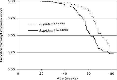
Low-penetrance breast cancer susceptibility alleles seem to play a significant role in breast cancer risk but are difficult to identify in human cohorts. A genetic screen of 176 N2 backcross progeny of two Trp53(+/-) strains, BALB/c and C57BL/6, which differ in their susceptibility to mammary tumors, identified a modifier of mammary tumor susceptibility in an approximately 25-Mb interval on mouse chromosome 7 (designated SuprMam1). Relative to heterozygotes, homozygosity for BALB/c alleles of SuprMam1 significantly decreased mammary tumor latency from 70.7 to 61.1 weeks and increased risk twofold (P = 0.002). Dmbt1 (deleted in malignant brain tumors 1) was identified as a candidate modifier gene within the SuprMam1 interval because it was differentially expressed in mammary tissues from BALB/c-Trp53(+/-) and C57BL/6-Trp53(+/-) mice. Dmbt1 mRNA and protein was reduced in mammary glands of the susceptible BALB/c mice. Immunohistochemical staining demonstrated that DMBT1 protein expression was also significantly reduced in normal breast tissue from women with breast cancer (staining score, 1.8; n = 46) compared with cancer-free controls (staining score, 3.9; n = 53; P < 0.0001). These experiments demonstrate the use of Trp53(+/-) mice as a sensitized background to screen for low-penetrance modifiers of cancer. The results identify a novel mammary tumor susceptibility locus in mice and support a role for DMBT1 in suppression of mammary tumors in both mice and women.


Figures

Figure 1
Figure 1
Linkage analysis results for mammary tumor susceptibility alleles. A: A complete genome scan was conducted comparing mammary tumor-bearing versus nonmammary tumor-bearing N2-Trp53+/− mice. Using 150 SNPs distributed across the mouse genome, significant linkage was detected on chromosome 7. B: Additional SNPs were used to define an interval of strong linkage on chromosome 7. The SuprMam1 locus was considered to span the region from 100 to 125 Mb. Locations of markers are based on the Ensembl mouse map, build 34.
Figure 2
Figure 2
Kaplan-Meier mammary tumor-free survival plots of N2-Trp53+/− mice segregated according to genotype at SuprMam1. Median age of occurrence of mammary tumors among mice that were heterozygous for the susceptibility allele (SuprMam1BALB/B6) was 70.7 weeks compared with 61.1 weeks for mice that were homozygous for the susceptibility alleles (SuprMam1BALB/BALB).
Figure 3
Figure 3
Expression of Dmbt1 in small intestines and mammary glands from BALB/c-Trp53+/− and C57BL/6-C57BL/6-p53+/− mouse strains. A: Northern blots were hybridized with a 32P-labeled probe from the 3′ end of the mouse Dmbt1 cDNA. The blots were stripped and rehybridized with a 32P-labeled Gapdh cDNA. B: The levels of Dmbt1 mRNA expression in small intestine and mammary gland were quantified using phosphor imaging and normalized relative to Gapdh to control for loading variations. Levels of Dmbt1 were significantly reduced in mammary tissue from BALB/c compared with C57BL/6 (*P = 0.002).
Figure 4
Figure 4
RT-PCR was used to analyze the structure of the 3′ end of Dmbt1 transcripts in mammary gland and small intestine. Top: The structure of DMBT1 protein and the location of the sequences of primers used for PCR. Bottom: Both strains of mice express sequences consistent with Dmbt1β. Only the levels of expression seem to differ between the strains.
Figure 5
Figure 5
Expression and localization of DMBT1 protein in mammary glands and small intestines. Tissues were collected from nulliparous BALB/c-Trp53+/− and C57BL/6-p53+/− mice and analyzed by immunohistochemistry. A: Punctate staining was present within the mammary epithelium of C57BL/6-p53+/− mice. Secretions were also evident within the lumen. B: The BALB/c-Trp53+/− mice were primarily devoid of staining in the mammary epithelium, but detectable levels were observed in some ducts as shown. In contrast, levels of expression were similar in the small intestines from both the C57BL/6-p53+/− (C) and BALB/c-Trp53+/− (D) strains. E: Expression of DMBT1 protein was examined in spontaneous mammary tumors in [C57BL/6 × BALB/c]-F1-Trp53+/− (F1) and BALB/c-Trp53+/− mice. Among the mammary tumors in F1 mice, there were equal numbers with significant expression as well as tumors with no detectable protein (i and ii, respectively). The mammary tumors from the BALB/c-Trp53+/− mice were primarily devoid of staining (iii) with only one that had very weak staining (iv).
Figure 6
Figure 6
Expression of DMBT1 mRNA in human tissues. A: Levels of DMBT1 expression were compared in normal human tissues using Q-PCR. Only lung expressed significant levels. The immortalized normal breast epithelial cells (76N series) also expressed high levels of DMBT1 mRNA. In contrast, expression of DMBT1 mRNA was undetectable in the primary breast cancers (BR18, BR19, BR20, BR21, BR24, BR25). NTC, no template control. All samples were analyzed for β-actin to ensure integrity of RNA. B: RT-PCR was used to analyze expression of DMBT1 mRNA in normal, immortalized breast epithelial cell lines (76N + Tert, 76N + 239, MCF10A), breast cancer cell lines (MCF-7, T47-D), and isogenic cells representing the primary breast tumor (21PT) and metastatic tumor (21MT) from a single patient. HeLa cells were used as a positive control. C: Immunohistochemistry for DMBT1 in benign human mammary epithelium. Immunohistochemical staining revealed variable patterns and intensities of staining among individuals. i: Diffuse cytoplasmic staining that was low intensity of reaction was considered score = 1. ii: Intense, focal expression within the supranuclear region and polarized toward the luminal surface was considered score = 2. iii: Intense staining polarized toward the luminal surface was considered score = 3.

References

    1. Lichtenstein P, Holm NV, Verkasalo PK, Iliadou A, Kaprio J, Koskenvuo M, Pukkala E, Skytthe A, Hemminki K. Environmental and heritable factors in the causation of cancer—analyses of cohorts of twins from Sweden, Denmark, and Finland. N Engl J Med. 2000;343:78–85. - PubMed
    1. Anglian Breast Cancer Study Group Prevalence and penetrance of BRCA1 and BRCA2 mutations in a population-based series of breast cancer cases. Anglian Breast Cancer Study Group. Br J Cancer. 2000;83:1301–1308. - PMC - PubMed
    1. Ford D, Easton DF, Stratton M, Narod S, Goldgar D, Devilee P, Bishop DT, Weber B, Lenoir G, Chang-Claude J, Sobol H, Teare MD, Struewing J, Arason A, Scherneck S, Peto J, Rebbeck TR, Tonin P, Neu-hausen S, Barkardottir R, Eyfjord J, Lynch H, Ponder BA, Gayther SA, Zelada-Hedman M, The Breast Cancer Linkage Consortiu Genetic heterogeneity and penetrance analysis of the BRCA1 and BRCA2 genes in breast cancer families. Am J Hum Genet. 1998;62:676–689. - PMC - PubMed
    1. Newman B, Mu H, Butler LM, Millikan RC, Moorman PG, King MC. Frequency of breast cancer attributable to BRCA1 in a population-based series of American women. JAMA. 1998;279:915–921. - PubMed
    1. Whittemore AS, Gong G, Itnyre J. Prevalence and contribution of BRCA1 mutations in breast cancer and ovarian cancer: results from three U.S. population-based case-control studies of ovarian cancer. Am J Hum Genet. 1997;60:496–504. - PMC - PubMed

Publication types

MeSH terms