. 2007 Jun;170(6):2112-21.
doi: 10.2353/ajpath.2007.060975.

Down-regulation of WAVE3, a metastasis promoter gene, inhibits invasion and metastasis of breast cancer cells

Affiliations

Down-regulation of WAVE3, a metastasis promoter gene, inhibits invasion and metastasis of breast cancer cells

Khalid Sossey-Alaoui et al. Am J Pathol. 2007 Jun.

Abstract

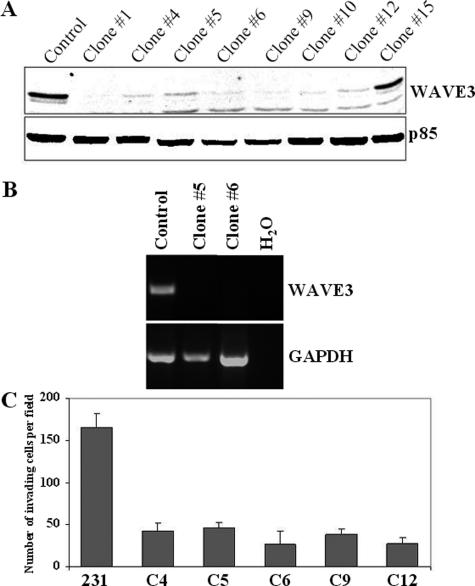
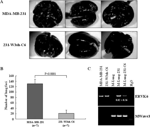
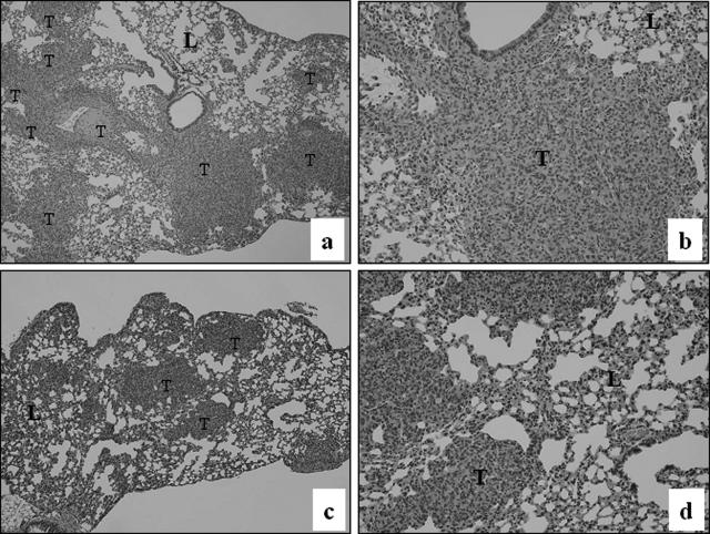
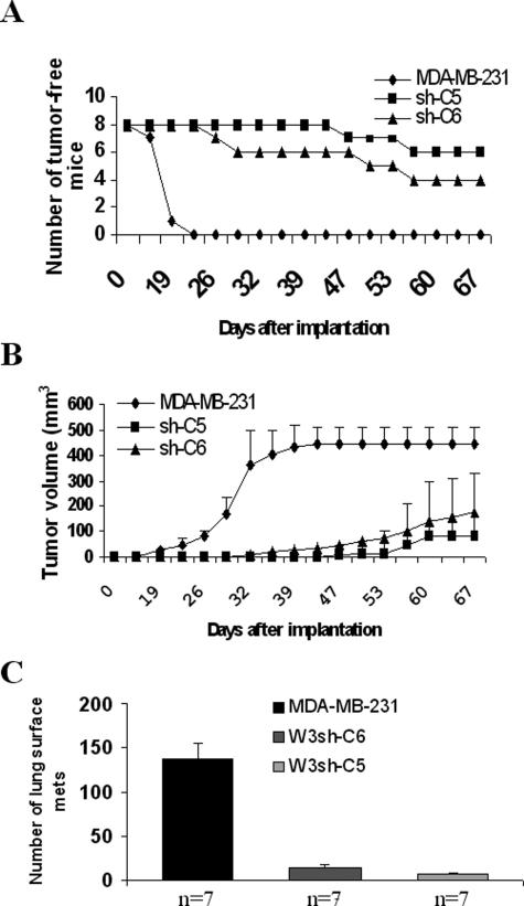
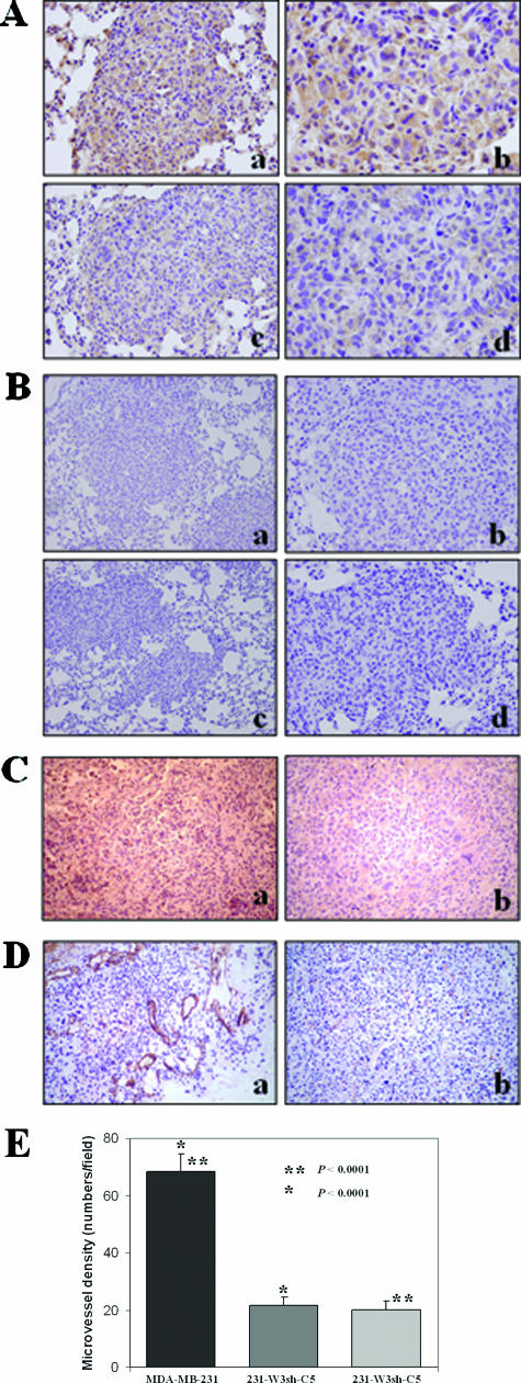
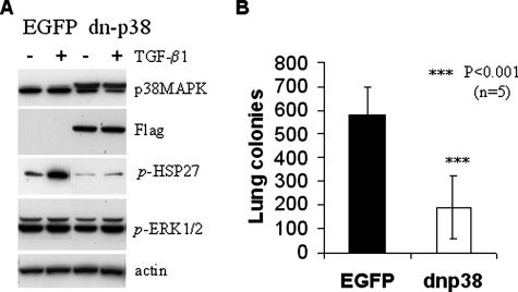
The expression of WAVE3, an actin-cytoskeleton and reorganization protein, is elevated in malignant human breast cancer, yet the role of WAVE3 in promoting tumor progression remains undefined. We have recently shown that knockdown of WAVE3 expression in human breast adenocarcinoma MDA-MB-231 cells using small interfering RNA resulted in a significant reduction of cell motility, migration, and invasion, which correlated with a reduction in the levels of active p38 mitogen-activated protein kinase. Here, we investigated the effect of stable suppression of WAVE3 by short hairpin RNA on tumor growth and metastasis in xenograft models. Breast cancer MDA-MB-231 cells expressing short hairpin RNA to WAVE3 (shWAVE3) showed a significant reduction in Matrigel invasion and formation of lung colonies after tail-vein injection in SCID mice. In the orthotopic model, we observed a reduction in growth rate of the primary tumors, as well as in the metastases to the lungs. We also show that suppression of p38 mitogen-activated protein kinase activity by dominant-negative p38 results in comparable phenotypes to the knockdown of WAVE3. These studies provide direct evidence that the WAVE3-p38 pathway contributes to breast cancer progression and metastasis.


Figures

Figure 1
Figure 1
Knockdown of WAVE3 expression with shRNA inhibits in vitro invasion of MDA-MB-231 cells. A: Western blot analysis of WAVE3 protein in independent clones derived from MDA-MB-231 cells with stable expression of shRNAs against WAVE3. WAVE3 protein levels were reduced to almost undetectable levels in all but clone 15. p85 protein was used and a loading control. B: RT-PCR analysis of WAVE3 mRNA in clones 5 and 6. WAVE3 mRNA expression was reduced to undetectable levels in clones 5 and 6. C: Results from the in vitro invasion assay shows the reduced ability of cells from independent clones with WAVE3 down-regulation to migrate through the Matrigel membrane. There was an average of fivefold reduction in invasion ability compared with the parental cells.
Figure 2
Figure 2
WAVE3 is overexpressed in human breast cancer. Immunohistochemical analysis of primary human breast cancer. In A, the top two panels (a, b) are from low-grade breast cancer showing weak staining for WAVE3 in the infiltrating tumor cells. The bottom two panels (c, d) are from a high-grade breast cancer showing a strong diffuse granular cytoplasmic pattern of staining in the infiltrating tumor cells. T, tumor; S, stromal tissue. WAVE3 staining in the tumors was scored as 0 for negative, 1 for weak, 2 for moderate, and 3 for strong. B: Comparison of the WAVE3 staining in the tissue sections demonstrates a significant difference between low- and high-grade tumors. The overall WAVE3 staining intensity was determined as the product of the percentage of positive cells and the staining score. Original magnifications: ×200 (a, c); ×400 (b, d).
Figure 3
Figure 3
Knockdown of WAVE3 expression inhibits in vivo metastasis in SCID mice. A: Pulmonary metastasis of MDA-MB-231 cells is suppressed by shRNA to WAVE3. A representative appearance of murine lungs with metastasized colonies (visible as white spots) from parental MDA-MB-231 cells, compared with the WAVE3-suppressed cells (clone 6 as an example), 5 weeks after intravenous injection. B: When the incidence of tumors on the surfaces of the lungs were counted in seven mice, the WAVE3-suppressed cells showed a significant decrease. C: PCR analysis of DNA from murine lungs after tail vein injection of shW3 clone 6 cells. The human-specific ERVK6A gene can be identified in human DNA (MDA-MB-231 and 231-shW3-C6) but not in mouse DNA (M-Lung). The presence of the human retroviral sequences in the mouse tissues (M-Lung-231 and M-Lung-231-C6) indicates the presence of human cells in these tissues. ERVK6A PCR amplification in M-Lung-231 relative to 231-shW3-C6 was calculated from the ratio of respective band intensities shown on the gel (fold ± SD from five independent PCR reactions). The mouse Wave3 gene is used as a control for integrity of the mouse DNA in each sample.
Figure 4
Figure 4
H&E staining of sections from lungs of mice injected with parental MDA-MB-231 cells (top) shows the presence of large tumors (a, b) compared with (bottom) lung metastases from the mice injected with cells expressing shWAVE3 (c, d). T, tumors; L, lung. Original magnifications: ×40 (a, c); ×100 (b, d).
Figure 5
Figure 5
Effect of WAVE3 knockdown on MDA-MB-231 tumor growth and lung metastasis in the spontaneous metastasis model. Tumor incidence and latency in mammary fat pads are shown in A, in which 100% of mice in the control group produced tumors, whereas the tumor incidence was reduced to 25 and 50% in the groups of mice implanted with clones W3sh-C5 and W3sh-C6, respectively. In B, the tumor growth curves represent the parental MDA-MB-231 cells, or C5 or C6, two clones derived from MDA-MB-231 cells expressing the shRNA against WAVE3, respectively. Each data point represents the mean of tumor volume ± SD, showing a significant difference (P < 0.00001) between the parental cells and the shWAVE3-derived cells. Mice were monitored for 67 days after implantation, after which they were all sacrificed and analyzed for lung metastases. C: Pulmonary metastasis is also suppressed by WAVE3 shRNA. The average number of lung metastases was ∼10 times higher in the control group compared with the shWAVE3 groups (P < 0.00001). n, number of mice per group.
Figure 6
Figure 6
Immunohistochemistry analysis of the tumors derived either from the parental MDA-MB-231 cells or from the WAVE3 knockdown cells in the subcutaneous model. A: Staining for the WAVE3 protein in the lung tumors from wild-type MDA-MB-231 cells show high levels of WAVE3 protein (a, b), compared with one of the rare lung metastases from cells expressing shWAVE3 (c, d), which shows marked reduction in WAVE3 levels, although some immunoreactivity is still seen. B: IgG staining as a negative control for the WAVE3 antibody in tumors from parental MDA-MB-231 (a, b) and of tumors from cells expressing shWAVE3 (c, d). C: Immunostaining for VEGF shows higher immunoreactivity in the MDA-MB-231-derived tumor (a), compared with the tumors derived from the WAVE knockdown cells (b). D: Immunostaining for CD31 demonstrates relatively large numbers of blood vessels and abnormal tumor vasculature in the MDA-MB-231-derived tumors (a), whereas fewer and more regular-shaped blood vessels are seen the WAVE knockdown-derived tumors (b). E: Microvessel density within tumor sections was determined in five fields of each tumor section, and presented as the mean number of microvessels per field. Original magnifications: ×100 (Aa, Ac, Bb, Bd); ×400 (Ab, Ad, E); ×40 (Ba, Bc).
Figure 7
Figure 7
Effect of a dominant-negative p38MAPK protein on MDA-MB-231 metastasis to the lungs. A: Immunoblot of whole cell extracts from cells expressing either the EGFP control or FLAG-tagged dn-p38 MAPK (dn-p38). The dn-p38 protein is associated with reduced phosphorylation of HSP27 but does not affect levels of phospho-activated ERK1/2. Cells were either untreated (−), or treated (+) with 2 ng/ml TGF-β1 for 0 to 24 hours as indicated. β-Actin was used as loading control. B: Colonization of lung surfaces by control EGFP-transfected MDA-MB-231 cells compared with cells expressing dn-p38 cells after injection into the lateral tail vein of SCID female mice (n, number of mice per group).

References

    1. Hanahan D, Weinberg RA. The hallmarks of cancer. Cell. 2000;100:57–70. - PubMed
    1. Friedl P, Wolf K. Tumour-cell invasion and migration: diversity and escape mechanisms. Nat Rev Cancer. 2003;3:362–374. - PubMed
    1. Billadeau DD. Cell growth and metastasis in pancreatic cancer: is Vav the Rho’d to activation? Int J Gastrointest Cancer. 2002;31:5–13. - PubMed
    1. Li Y, Tondravi M, Liu J, Smith E, Haudenschild CC, Kaczmarek M, Zhan X. Cortactin potentiates bone metastasis of breast cancer cells. Cancer Res. 2001;61:6906–6911. - PubMed
    1. Michiels F, Habets GG, Stam JC, van der Kammen RA, Collard JG. A role for Rac in Tiam1-induced membrane ruffling and invasion. Nature. 1995;375:338–340. - PubMed

Publication types

Substances