. 2007 May 25;316(5828):1194-8.
doi: 10.1126/science.1139476.

Abraxas and RAP80 form a BRCA1 protein complex required for the DNA damage response

Affiliations

Abraxas and RAP80 form a BRCA1 protein complex required for the DNA damage response

Bin Wang et al. Science. .

Abstract

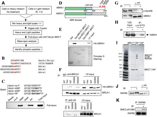
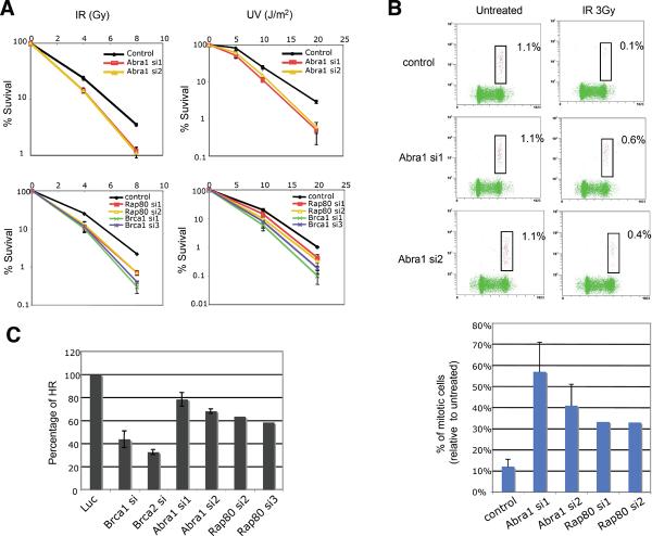
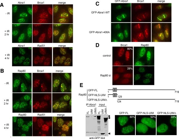
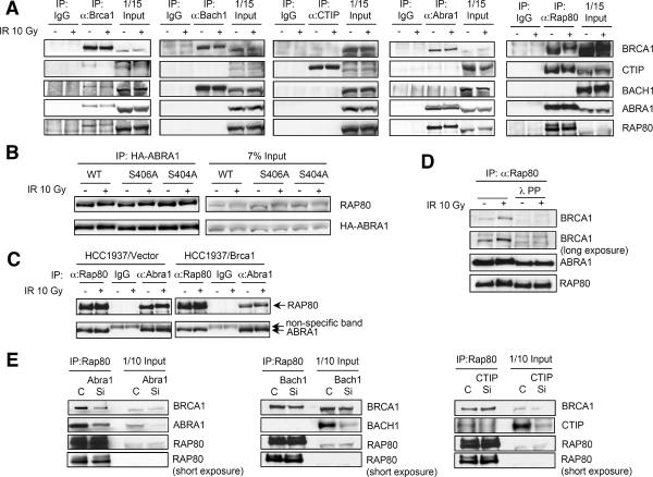
The BRCT repeats of the breast and ovarian cancer predisposition protein BRCA1 are essential for tumor suppression. Phosphopeptide affinity proteomic analysis identified a protein, Abraxas, that directly binds the BRCA1 BRCT repeats through a phospho-Ser-X-X-Phe motif. Abraxas binds BRCA1 to the mutual exclusion of BACH1 (BRCA1-associated C-terminal helicase) and CtIP (CtBP-interacting protein), forming a third type of BRCA1 complex. Abraxas recruits the ubiquitin-interacting motif (UIM)-containing protein RAP80 to BRCA1. Both Abraxas and RAP80 were required for DNA damage resistance, G(2)-M checkpoint control, and DNA repair. RAP80 was required for optimal accumulation of BRCA1 on damaged DNA (foci) in response to ionizing radiation, and the UIM domains alone were capable of foci formation. The RAP80-Abraxas complex may help recruit BRCA1 to DNA damage sites in part through recognition of ubiquitinated proteins.


Figures

Fig. 1
Fig. 1. Identification of Abraxas and Rap80 as Brca1-BRCT interacting proteins
(A) A schematic view of experimental procedures for identifying phospho-peptides bound to Brca1-BRCT. (B) Identified phospho-peptides sequences. The # symbol indicates a phosphate resides on the previous residue (C). Phosphorylated Abra1 peptides bind to purified recombinant Brca1 BRCT domains. Biotinylated peptides on streptavidin beads were used to pull down purified recombinant GST-Brca1-BRCT and visualized by Comassie staining. (D) Diagram of Abra1 and its paralog Abro1 protein structures. (E) Abraxas specifically binds to Brca1 BRCT domains. HA-Abra1 was expressed in 293T cells and cell lysates were incubated with different purified GST-tagged BRCT domains. (F) HA-Abra1 association with endogenous Brca1 is dependent on S406 phosphorylation. HA-Abra1 wild type or mutant proteins were expressed in 293T cells. (G) Ser406 of Abra1 is phosphorylated in vivo. Lysates from 293T cells treated or not treated with IR and lambda phosphatase were resolved by SDS-PAGE. (H) Abraxas can be recognized by phospho-SQ/TQ antibodies against ATM/ATR substrates. Lysates from 293T cells were immunoprecipitated with anti-phospho-S/TQ antibodies and Western blots were probed with anti-Abra1 antibodies. (I) Rap80 was identified in TAP purification of Brca1-BRCT domain associated proteins. Retroviruses expressing either TAP only (TAP) or C-terminal TAP-tagged BRCT domain of Brca1 (BRCT-TAP) were introduced into Hela cells, and the infected cells were used for purification. A coomassie stained gel is shown. (J) Rap80 is phosphorylated in response to IR. (K) Rap80 can be recognized by phospho-antibodies against ATM/ATR substrates. Lysates from 293T cells were immunoprecipitated with anti-Rap80 antibody and probed with the indicated antibody.
Fig. 2
Fig. 2. Abra1 and Rap80 are involved in DNA damage responses
(A) Abra1-depleted or Rap80-depleted cells are sensitive to IR and UV. U2OS cells were treated with control oligos or siRNAs against Abra1, Rap80 or Brca1, incubated for 2 days, plated at low density, irradiated, and colonies counted after 14 days. (B) Analysis of the G2/M checkpoint. Cells were untreated or treated with 3 Gy IR as indicated, then incubated for 1 h at 37°C before fixation and p-H3 antibody staining. Three independent experiments were performed with siRNA oligos against Abra1. Two independent experiments were performed with siRNA oligos against Rap80 that yielded similar results. (C) Abra1-siRNA treated cells or Rap80-siRNA treated cells are defective for homologous recombination. DR-U2OS cells were transfected with siRNAs against luciferase, Brca1, Brca2, Abra1 or Rap80 separately. siRNAs against Brca1 or Brca2 were a mixture of three different siRNA oligos for each gene. Individual siRNA oligos were used for Abra1 or Rap80 genes. Three independent experiments were performed with siRNAs against luciferease, Brca1, Brca2 and Abra1.
Fig. 3
Fig. 3. Abra1 and Rap80 form DNA damage induced foci
(A) and (B) Abra1 and Rap80 form DNA damage induced foci that colocalize with Brca1 and Rad51 foci. U2OS cells were either untreated or treated with IR 10 Gy for 2h, fixed and co-immunostatined with rabbit anti-Abra1 antibody, mouse anti-Brca1 antibody, or mouse anti-Rad51 antibody followed by proper Alex 488-conjugated or Cy3-conjuated antibodies. (C) Abra1 localizes to DNA damage regions as early as 15 min after damage, independent of binding to Brca1. Laser microirradiation was performed on U2OS cells carrying a stably integrated retroviral construct expressing GFP tagged wild type or mutant Abra1 (S406A). Cells were fixed and stained 15 min after laser treatment. (D) Brca1 foci formation is defective in Rap80-siRNA-treated cells. U2OS cells were transfected with control oligos or siRNAs against Rap80 for two days. Cells were then irradiated with 10 Gy IR, incubated for 2 h, fixed and immunostained with indicated antibodies. More than 400 cells were counted and cells containing more than 10 Brca1 foci were counted as positive. Three different oligos against Rap80 were used and similar results were obtained. (E) Rap80 IRIF formation is dependent on UIM domains independently of Abraxas binding. U2OS cells with stably integrated retroviral construct expressing GFP-WT, GFP-UIM or GFP-UIMΔ were irradiated with 10 Gy IR, 2h later, fixed and stained with antibodies against GFP followed with Alexa 488 secondary antibodies. More than 300 cells were counted to determine the percentage of cells forming foci for each cell line. Immunoprecipitation of Abraxas revealed binding to the C-terminal region of Rap80 lacking the UIM domain.
Fig. 4
Fig. 4. Abra1 and Rap80 exist in a complex with Brca1
(A) Brca1 forms distinct complexes with Abra1, Bach1 and CTIP. Immunoprecipitation with antibodies against Brca1, Bach1, CTIP, Abra1 and Rap80 was carried out with lysates from 293T cells treated or untreated with IR. (B) Rap80 interacts with the Abra1 mutant (S406A) that does not bind to Brca1. HA-tagged wild type or a mutant of Abra1 was expressed in 293T cells. Lysates of cells treated or untreated with IR were immunoprecipitated with anti-Rap80 antibodies. (C) TheRap80-Abra1 interaction is intact in HCC1937 cells that lack a functional Brca1. HCC1937 cells were either treated or untreated with IR. Cell lysates were immunoprecipitated with anti-Rap80, anti-Abra1 or control IgG. (D) The Rap80-Abra1 interaction is not phosphorylation dependent. Immunoprecipitation with Rap80 antibodies was carried out in cell lysates treated or not treated with lambda phosphatase. (E) The Rap80-Brca1 interaction decreases in cells treated with siRNA against Abra1. 293T cells were transfected with control oligo (C) or siRNA oligos against Abra1, Bach1 or CTIP (Si). 48 hrs later, cell lysates were prepared and immunoprecipitated with antibodies against Rap80. Immunoblotting was carried out with antibodies against Brca1, Abra1, Bach1, CTIP or Rap80.

Comment in

References

    1. Venkitaraman AR. Cell. 2002;108:171. - PubMed
    1. Narod SA, Foulkes WD. Nat Rev Cancer. 2004;4:665. - PubMed
    1. Cortez D, Wang Y, Qin J, Elledge SJ. Science. 1999;286:1162. - PubMed
    1. Wu LC, et al. Nat Genet. 1996;14:430. - PubMed
    1. Baer R, Ludwig T. Curr Opin Genet Dev. 2002;12:86. - PubMed

Publication types

Associated data