. 2007 May 25;316(5828):1198-202.
doi: 10.1126/science.1139516.

RAP80 targets BRCA1 to specific ubiquitin structures at DNA damage sites

Affiliations

RAP80 targets BRCA1 to specific ubiquitin structures at DNA damage sites

Bijan Sobhian et al. Science. .

Abstract

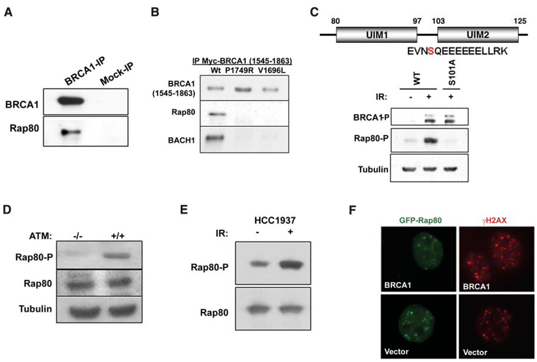
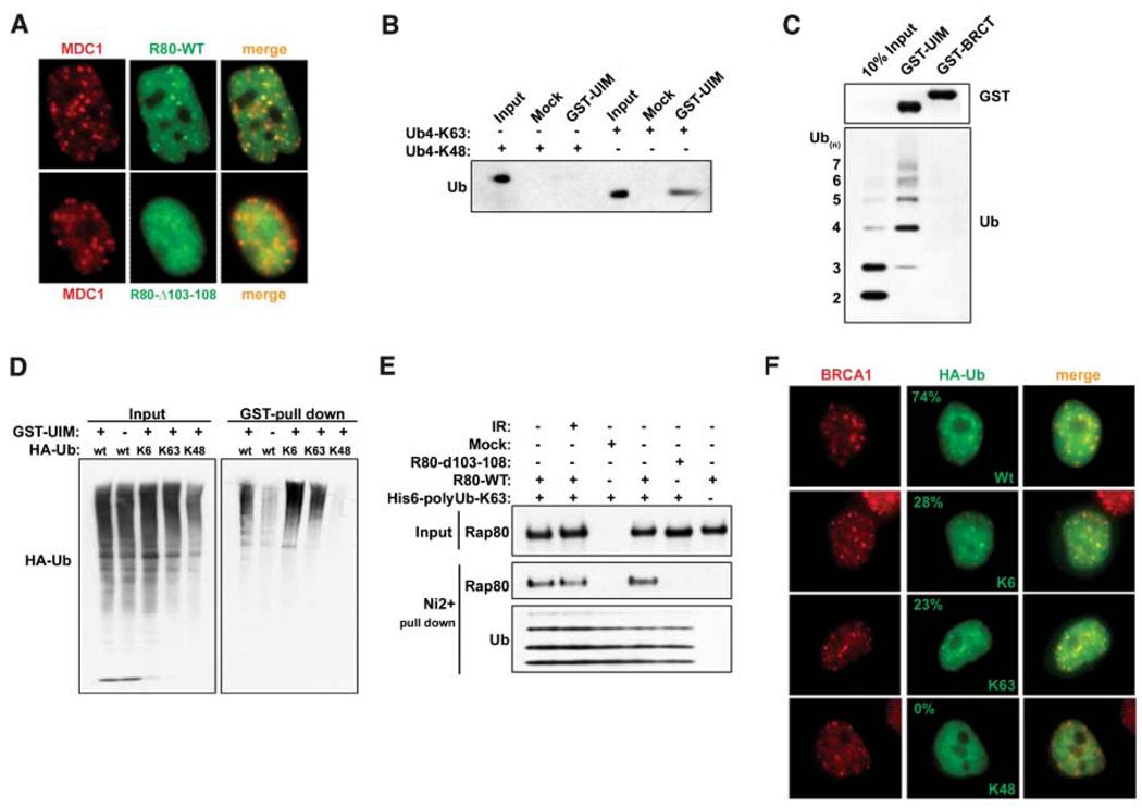
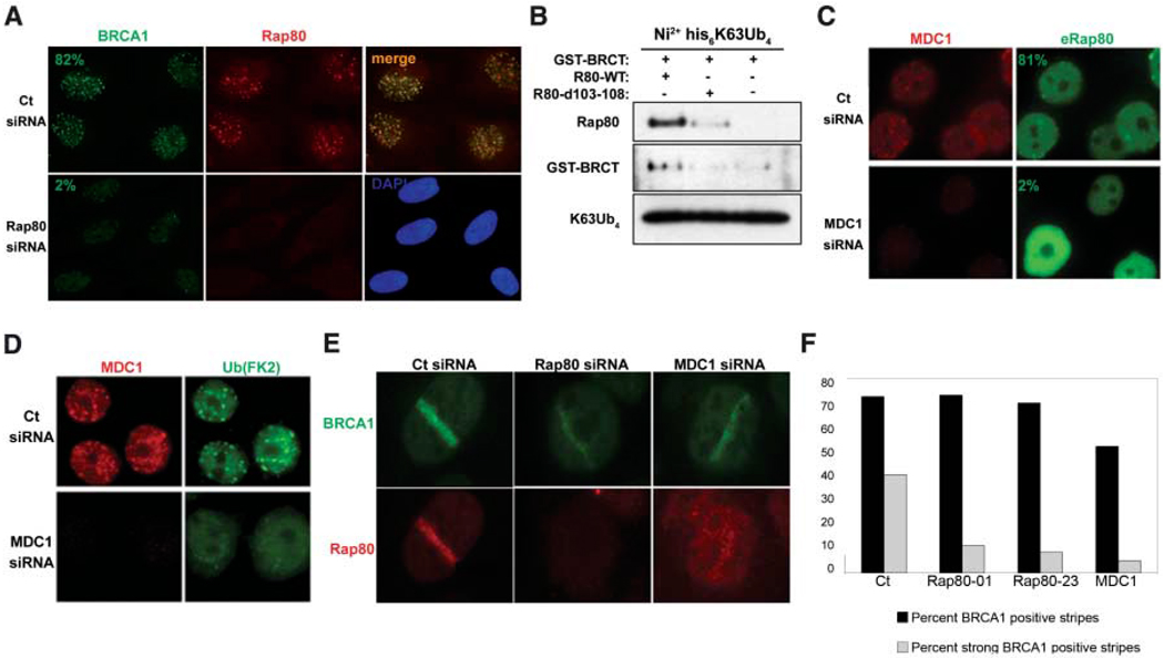
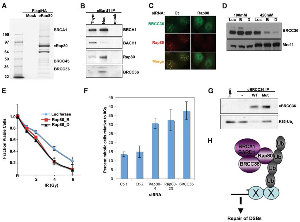
Mutations affecting the BRCT domains of the breast cancer-associated tumor suppressor BRCA1 disrupt the recruitment of this protein to DNA double-strand breaks (DSBs). The molecular structures at DSBs recognized by BRCA1 are presently unknown. We report the interaction of the BRCA1 BRCT domain with RAP80, a ubiquitin-binding protein. RAP80 targets a complex containing the BRCA1-BARD1 (BRCA1-associated ring domain protein 1) E3 ligase and the deubiquitinating enzyme (DUB) BRCC36 to MDC1-gammaH2AX-dependent lysine(6)- and lysine(63)-linked ubiquitin polymers at DSBs. These events are required for cell cycle checkpoint and repair responses to ionizing radiation, implicating ubiquitin chain recognition and turnover in the BRCA1-mediated repair of DSBs.


Figures

Fig. 1
Fig. 1
RAP80 interacts with the BRCA1 BRCT motif and responds to IR independently of BRCA1. (A) Co-immunoprecipitation (IP) between endogenous BRCA1 and RAP80. (B) Lysates were prepared from 293T cells transfected with WT or clinical mutant myc-BRCA1-BRCT domains. Myc antibody immunoprecipitated material was separated by SDS–polyacrylamide gel electrophoresis, and immunoblotting (IB) was performed. (C) HeLa S3 cells stably expressing epitope tagged-WT or S101A mutant RAP80 were IR-treated, and lysates were probed with an antibody specific to phosphorylated (P) RAP80-S-101. E, Glu; L, Leu; N, Asn; R, Arg; and V, Val. (D) ATM −/− and +/+ and (E) HCC1937 cells were gamma irradiated, and IB was performed on cell lysates as indicated. (F) HCC1937 cells reconstituted with vector or WT BRCA1 were transfected with GFP-RAP80 and gamma-irradiated, and, 1 hour later, IF was performed to assess colocalization with γH2AX.
Fig. 2
Fig. 2
RAP80 forms IRIF by binding to non-K48–linked ubiquitin. (A) HeLa cells stably expressing FLAG-HA– tagged WT or Δ103–108 RAP80 were treated with 6 Gy IR, and IF was performed 1 hour later. (B) K63- or K48-linked tetraubiquitin was incubated with the RAP80 GST-UIM domain. GST precipitations were analyzed by IB. (C) A mixture of K63-linked ubiquitin polymers containing two to seven molecules of ubiquitin (Ub2–7) was incubated with RAP80 GST-UIM or GST-BRCT. IB was performed as indicated after purification on glutathione-conjugated sepharose beads. (D) 293T cells transfected with the indicated HA- tagged ubiquitin expression vectors were treated with 10 µM MG132 for 2 hours before lysis and then incubated with RAP80 GST-UIM protein. The bound ubiquitin species were analyzed by IB. (E) FLAG-HA–tagged WT or Δ103–108 RAP80 were purified before and after IR from stably expressing HeLa-S3 cells by FLAG IP followed by FLAG-peptide elution. These proteins were then incubated with His-tagged, K63-Ub2–7, followed by purification on Ni2+- agarose beads. Ubiquitin-associated proteins were detected by IB. (F) HeLa cells were transfected with the same ubiquitin-expression vectors as in (D) and analyzed by IF 1 hour after IR. The percentage of transfected cells that display colocalization of epitope-tagged ubiquitin and BRCA1 is indicated (N > 200).
Fig. 3
Fig. 3
RAP80 targets BRCA1 to MDC1-dependent polyubiquitin structures at DSBs. (A) U2OS cells transfected with control (Ct) or RAP80-specific siRNA were fixed 6 hours after 10 Gy IR and analyzed by IF. (B) FLAG IP-purified WT or Δ103–108 RAP80 derived from HeLa S3 cells stably expressing these proteins was incubated with GST-BRCT and K63-linked tetraubiquitin. Ni2+-agarose precipitations were analyzed by IB as indicated. (C and D) HeLa cells transfected with Ct or MDC1-specific siRNA were fixed 6 hours after 6 Gy IR and analyzed by IF. (E) U2OS cells treated with the indicated specific siRNA were fixed at either 15 min or 4 hours after microirradiation by using a 337-nm laser and analyzed by IF. (F) Detection of the frequency and qualitative intensity of BRCA1 at γH2AX-positive stripes after treatment of U2OS cells with the indicated siRNA. Experiments were performed in duplicate at 15 min and 4 hours after laser-induced damage, and more than 125 stripes were counted per sample.
Fig. 4
Fig. 4
Interactions of RAP80, BRCC36, and BRCA1. (A) Coommassiestained gel of FLAG and HA double-purified RAP80 complexes from HeLa S3 cells stably expressing eRAP80. (B) HeLa S3 cells stably expressing eBARD1 were treated with 2 mM thymidine (Thym) or 0.5 µM nocodazole (Noc). FLAG-purified proteins were analyzed by IB. (C) U2OS cells expressing HA-tagged BRCC36 were transfected with shRNA specific to RAP80 or control and, 48 hours later, were monitored for IRIF at 1 hour after 6 Gy. (D) HeLa cells expressing stable siRNA vectors specific to luciferase (Luc) or RAP80 (B and D) were sequentially extracted with NETN buffer containing 100 mM NaCl and then NETN buffer containing 420 mM NaCl. Extracts were analyzed by IB. (E) HeLa cells expressing stable shRNA vectors specific to luciferase or RAP80 (B and D) were treated with the indicated doses of IR, and the number of viable cells were counted 96 hours later. The fraction of viable cells compared with a culture receiving 0 Gy is expressed graphically. Experiments were done in triplicate. Error bars indicate standard deviation. (F) The percentage of phosphohistone H3– positive cells (mitotic population) at 1 hour after 2 Gy IR compared with at 0 Gy (i.e., mock irradiation) was plotted for each siRNA-transfected U2OS population. Experiments were done in triplicate; error bars indicate standard deviation. (G) FLAG peptide eluates from FLAG IPs performed on extracts of HeLa S3 cells stably expressing FLAG-HA–tagged WT or a catalytically inactive BRCC36 mutant were incubated with K63-linked tetraubiquitin (K63-Ub4) for 30 min at 37°C. The reaction mixtures were then analyzed by IB. (H) Model for recruitment of a BRCA1-BARD1-BRCC36-RAP80 complex to sites of DNA damage by binding to non-K48–linked ubiquitin structures.

Comment in

References

    1. Bassing CH, et al. Cell. 2003;114:359. - PubMed
    1. Celeste A, et al. Cell. 2003;114:371. - PMC - PubMed
    1. Wang Y, et al. Nat. Genet. 2005;37:750. - PubMed
    1. Stewart GS, Wang B, Bignell CR, Taylor AM, Elledge SJ. Nature. 2003;421:961. - PubMed
    1. Stucki M, et al. Cell. 2005;123:1213. - PubMed

MeSH terms