. 2007 Jun 22;100(12):1732-40.
doi: 10.1161/CIRCRESAHA.107.148791. Epub 2007 May 24.

Enhancing repair of the mammalian heart

Affiliations

Enhancing repair of the mammalian heart

Maria Paola Santini et al. Circ Res. .

Abstract

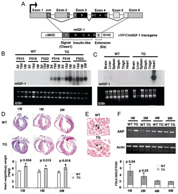
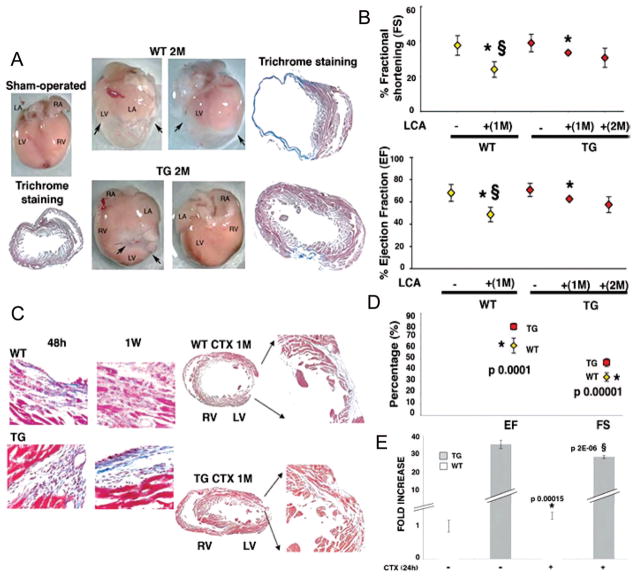
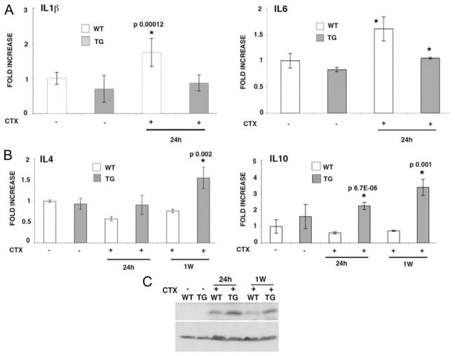
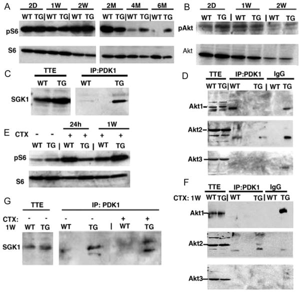
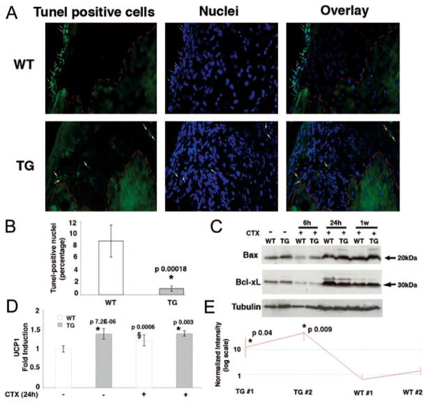
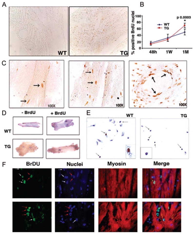
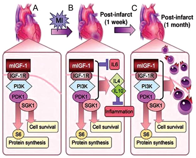
The injured mammalian heart is particularly susceptible to tissue deterioration, scarring, and loss of contractile function in response to trauma or sustained disease. We tested the ability of a locally acting insulin-like growth factor-1 isoform (mIGF-1) to recover heart functionality, expressing the transgene in the mouse myocardium to exclude endocrine effects on other tissues. supplemental mIGF-1 expression did not perturb normal cardiac growth and physiology. Restoration of cardiac function in post-infarct mIGF-1 transgenic mice was facilitated by modulation of the inflammatory response and increased antiapoptotic signaling. mIGF-1 ventricular tissue exhibited increased proliferative activity several weeks after injury. The canonical signaling pathway involving Akt, mTOR, and p70S6 kinase was not induced in mIGF-1 hearts, which instead activated alternate PDK1 and SGK1 signaling intermediates. The robust response achieved with the mIGF-1 isoform provides a mechanistic basis for clinically feasible therapeutic strategies for improving the outcome of heart disease.


Figures

Figure 1
Figure 1
Characterization of αMyHC/mIGF-1 transgenic mice. A, Schematic representation of the rodent Igf-1 gene. B and C, Northern blot analysis of total RNA from wild-type (WT) and transgenic (mIGF-1) hearts at different ages (B) and different tissues (C) using a rat Igf-1 32P labeled probe. Ethidium bromide (EtBr) was used to verify equal RNA loading. D, Histological analysis of WT and mIGF-1 transgenic (TG) hearts by Hematoxylin and Eosin staining. Lower panel shows adult heart weight/body weight (P<0.05). Values are the average of 6 independent analyses. E, Cell size differences in WT and TG hearts. F, RT-PCR analysis of the hypertrophic marker ANP in adult hearts. PCR values were normalized for β-actin content. Densitometric analysis was performed on 3 independent experiments. Asterisks indicate significant relative values (P<0.05).
Figure 2
Figure 2
Enhanced cardiac repair and functions in mIGF-1 transgenic mice after myocardial infarction. A, Whole mount and histological analysis of sham-operated control (WT) heart (left) and LCA WT and TG hearts (right) 2 months after operation. Arrows indicate fibrotic tissue. LA indicates left atrium; LV, left ventricle; RA, right atrium; RV, right ventricle. Histological analysis by trichrome staining is shown for each treatment. B, Functional recovery of mIGF-1 transgenic mice after LCA. Mean percentage values of FS (upper panel) and EF (lower panel) are representative of 3 readings on each animal and averaged among groups. Asterisks (*) show significant values (P<0.05) between uninjured and injured hearts in WT (yellow square) and TG (red square) mice. § shows significant values between WT and TG injured hearts. C, Histological analysis (trichrome) of WT and TG hearts at 48 hours, 1 week, and 1 month after CTX injection in the left ventricular wall. Comparable results were obtained with similar analyses on 6 different groups. D, Functional recovery of mIGF-1 transgenic mice 1 month after CTX injection. Mean percentage values are representative of 3 readings on each animal and averaged among groups. Asterisk (*) indicates significant values decreasing in WT compared with TG hearts (P<0.05). E, Real time PCR of mIGF-1 transcript in physiological conditions and 24 hours after CTX injection, using IGF-1Ea Taqman probe (Applied Biosystem). PCR values were normalized for GAPDH content in each sample. Asterisk (*) indicates significant increasing values compared with WT uninjured hearts, whereas § represents significant decreasing values compared with TG uninjured hearts.
Figure 3
Figure 3
Inflammatory markers are repressed early in mIGF-1–induced cardiac amelioration. A, Real-time PCR analysis of the inflammatory interleukins IL6 and IL1β 24 hours after CTX injection in WT and TG hearts. Real-time PCR was normalized by GAPDH content in each sample. In the left and right panels asterisks indicate significant increasing values compared with uninjured WT or TG hearts in 3 independent experiments. B, Real-time PCR analysis of the antiinflammatory cytokines IL4 and IL10 in TG and WT hearts 24 hours and 1 week after CTX injection. PCR was normalized by GAPDH content in each sample. C, Western blot analysis of p21 protein content in WT and TG hearts 24 hours and 1 week after CTX injection. β-actin was used to normalize equal protein loading.
Figure 4
Figure 4
mIGF-1 induces interaction of PDK1 with SGK1 but not with Akt to phosphorylate S6. A and B, Western blot analysis of Akt and S6 ribosomal protein phosphorylation. Each analysis is representative of 3 independent experiments with no significant variation (data not shown). C andD, IP-Western analysis of PDK1 interaction with SGK1 and Akt isoforms. No PDK1/Akt interaction was observed. TTE indicates total tissue extract. Additional bands present in Akt1 and Akt2 blots are attributable to antibody cross-reaction and to nonspecific interaction with IgG. E, Western blot analysis of S6 ribosomal protein phos-phorylation after CTX cardiac injury. Note persistence of pS6 levels in TG hearts at 1 week after injury. F and G, IP-Western analysis of PDK1 interaction with SGK1 and Akt isoforms after CTX cardiac injury. No PDK1/Akt interaction was observed.
Figure 5
Figure 5
mIGF-1 transgenic expression protects against DNA damage and increases expression of the mitochondrial protein UCP1. A, TUNEL assay analysis of WT (upper panel) and TG hearts (lower panels) 1 week after CTX injection. White arrows in the WT heart indicate TUNEL-positive nuclei, whereas the TG heart showed a nonspecific signal. Red outline demarcates the border of the injury. B, Percentage amount of TUNEL-positive cells 1 week after CTX injection. Asterisk (*) indicates a significant decrease of apoptotic cells in TG hearts. C, Expression analysis of the pro-apoptotic markers Bax and Bcl-xL 6 hours, 24 hours, and 1 week after CTX injection. D, UCP1 RT-PCR of uninjured WT and TG hearts and injured WT and TG hearts 24 hours after CTX injection. Asterisk indicates a significant increase of UCP1 transcripts in TG hearts compared with WT hearts with or without CTX-induced infarct. § indicates UCP1 increasing levels compared with uninjured hearts. E, Affymetrix analysis of UCP1. The transcript is expressed as logarithmic scale of intensity in 2 TG and WT hearts. * indicates a significant increase of UCP1 transcripts in TG hearts.
Figure 6
Figure 6
Late cell proliferation in mIGF-1 transgenic hearts. A, Positive nuclei in paraffin sections visualized with biotinylated anti-BrdU antibody in the hearts of WT and TG mice. B, Statistical analysis of BrdU-positive nuclei in paraffin sections stained with biotinylated anti-BrdU antibody and counted at different time points after CTX injection. Asterisk indicates significant relative values in the BrdU-positive hearts at 1 month (P<0.05). Values are the average of 3 independent experiments. C, Characteristics of BrdU-positive cells in mIGF-1 hearts 1 month after CTX injection. BrdU-positive cells were photographed at 100× magnification. Arrows indicate BrdU-positive cardiomyocytes (left and middle panels) and cells lining blood vessel (right panel). Cardiac myocytes (D) and nonmuscle cells (E) were isolated from BrdU-labeled WT and TG hearts 1 month after CTX injection. Dissociated cell cultures were analyzed for BrdU and hematoxylin to visualize proliferating nuclei. The experiment was performed on 3 hearts each from WT and TG mice. F, Confocal microscopic analysis of BrdU-positive cells in TG heart tissue at 100× magnification. Cardiomyocytes were visualized by an anti-myosin antibody. White arrows indicate BrdU-positive cardiac myocytes; red arrows indicate noncardiomyocyte cells.
Figure 7
Figure 7
Mechanisms of mIGF-1 induce recovery in injured hearts. A, Transgenic mIGF-1 induces protein synthetic and cell survival pathways in cardiac tissue through a PDK1/SGK1 phosphorylation cascade, bypassing canonical Akt, mTOR, and p70S6K intermediates. B, On myocardial infarction (MI), pathways induced by mIGF-1 result in rapid repression of proinflammatory cytokines such as IL6 that promote fibrosis and cardiac decompensation while activating cytokines such as IL4 and IL10 that resolve inflammation. C, This permissive tissue environment enables efficient cardiac wall replacement, as shown by increased proliferation of cells at 1 month after injury, and functional repair in mIGF-1 transgenic hearts.

References

    1. Longo VD, Finch CE. Evolutionary medicine: from dwarf model systems to healthy centenarians? Science. 2003;299:1342–1346. - PubMed
    1. Adamo ML, Neuenschwander S, LeRoith D, Roberts CT., Jr Structure, expression, and regulation of the IGF-I gene. Adv Exp Med Biol. 1993;343:1–11. - PubMed
    1. Goldspink G. Gene expression in skeletal muscle. Biochem Soc Trans. 2002;30:285–290. - PubMed
    1. Chan JM, Stampfer MJ, Giovannucci E, Gann PH, Ma J, Wilkinson P, Hennekens CH, Pollak M. Plasma insulin-like growth factor-I and prostate cancer risk: a prospective study. Science. 1998;279:563–566. - PubMed
    1. Reiss K, Cheng W, Ferber A, Kajstura J, Li P, Li B, Olivetti G, Homcy CJ, Baserga R, Anversa P. Overexpression of insulin-like growth factor-1 in the heart is coupled with myocyte proliferation in transgenic mice. Proc Natl Acad Sci U S A. 1996;93:8630–8635. - PMC - PubMed

Publication types

MeSH terms